 Explore
 Validate
 Learn
Learn Western blot
 Immunocytochemistry
 Immunoprecipitation
Antibody data
- Antibody Data
- Antigen structure
- References [4]
- Comments [0]
- Validations
- Immunocytochemistry [2]
Submit
Validation data
Reference
Comment
Report error
- Product number
- ALX-804-057-C100 - Provider product page

- Provider
- Enzo Life Sciences
- Proper citation
- Enzo Life Sciences Cat#ALX-804-057-C100, RRID:AB_2052202
- Product name
- Perforin (mouse) monoclonal antibody (CB5.4)
- Antibody type
- Monoclonal
- Antigen
- Recombinant protein fragment
- Reactivity
- Mouse
- Host
- Rat
- Isotype
- IgG
- Antibody clone number
- CB5.4
- Vial size
- 100 μg
- Storage
- -20°C
- Handling
- Avoid freeze/thaw cycles.
Submitted references Fas-Fas ligand interactions are essential for the binding to and killing of activated macrophages by gamma delta T cells.
Cubilin, a binding partner for galectin-3 in the murine utero-placental complex.
Resistance of CTL to perforin-mediated lysis. Evidence for a lymphocyte membrane protein interacting with perforin.
The calcium-binding protein calreticulin is a major constituent of lytic granules in cytolytic T lymphocytes.
Dalton JE, Howell G, Pearson J, Scott P, Carding SR
Journal of immunology (Baltimore, Md. : 1950) 2004 Sep 15;173(6):3660-7
Journal of immunology (Baltimore, Md. : 1950) 2004 Sep 15;173(6):3660-7
Cubilin, a binding partner for galectin-3 in the murine utero-placental complex.
Crider-Pirkle S, Billingsley P, Faust C, Hardy DM, Lee V, Weitlauf H
The Journal of biological chemistry 2002 May 3;277(18):15904-12
The Journal of biological chemistry 2002 May 3;277(18):15904-12
Resistance of CTL to perforin-mediated lysis. Evidence for a lymphocyte membrane protein interacting with perforin.
Müller C, Tschopp J
Journal of immunology (Baltimore, Md. : 1950) 1994 Sep 15;153(6):2470-8
Journal of immunology (Baltimore, Md. : 1950) 1994 Sep 15;153(6):2470-8
The calcium-binding protein calreticulin is a major constituent of lytic granules in cytolytic T lymphocytes.
Dupuis M, Schaerer E, Krause KH, Tschopp J
The Journal of experimental medicine 1993 Jan 1;177(1):1-7
The Journal of experimental medicine 1993 Jan 1;177(1):1-7
No comments: Submit comment
Supportive validation
- Submitted by
- Enzo Life Sciences (provider)
- Main image

- Experimental details
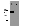
- Detection of perforin in the mouse T cell clone B6.1. (lane 1). Absence of perforin in the mouse fibroblast cell line NIH.3T3 (lane 2). The protein migrates as a 66 kDa species. Method: Cell extracts from the T cell clone B6.1 (2x10E6) were resolved by SDS-PAGE under reducing conditions, transferred to nitrocellulose and probed with the CB5.4 antibody at 1 µg/ml. Proteins were visualized using a peroxidase-conjugated antibody to rat IgG and a chemiluminescence detection system.
- Submitted by
- Enzo Life Sciences (provider)
- Main image
- Experimental details
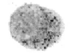
- Detection of perforin in an individual mouse T cell. Perforin is located in cytoplasmic granules. Method: A mouse T cell clone was plated onto polylysine treated glass slides, fixed and permeabilized in methanol at -20oC for 5 min, then in acetone at -20oC for 30 sec. After 3 washings in PBS, 0.1% BSA, slides were incubated with 5 µg/ml of CB5.4 antibody, washed in PBS, incubated in anti-rat IgG-HRP, washed in PBS, and developed in appropriate substrate solution (DAB or AEC).
