 Explore
 Validate
 Learn
Learn Western blot
 ELISA
ELISAAntibody data
- Antibody Data
- Antigen structure
- References [0]
- Comments [0]
- Validations
- Western blot [1]
- Immunohistochemistry [2]
Submit
Validation data
Reference
Comment
Report error
- Product number
- AP09263PU-N - Provider product page

- Provider
- Acris Antibodies GmbH
- Proper citation
- Acris Antibodies GmbH Cat#AP09263PU-N, RRID:AB_2035980
- Product name
- anti Sprouty homolog 4 / SPRY4 (306-322)
- Antibody type
- Polyclonal
- Antigen
- Synthetic peptide corresponding to amino acids 306-322 of Human Sprouty-4 protein
- Reactivity
- Human, Mouse, Rat, Bovine, Canine
- Host
- Rabbit
- Isotype
- IgG
- Vial size
- 0.1 mg
- Concentration
- 0.93 mg/ml (by UV absorbance at 280 nm)
No comments: Submit comment
Supportive validation
- Submitted by
- Acris Antibodies GmbH (provider)
- Main image

- Experimental details
- Western blot using anti-Sprouty-4 antibody shows detection of a doublet band ~35 kDa corresponding to human Sprouty-4. Approximately 30 μg of HeLa (lane 1), SW13 (lane 2) and C2C12 (Lane 3) whole cell lysates were separated by 10% SDS-PAGE and transferred ontonitrocellulose. After blocking with 5% non-fat dry milk in TBST the membrane was probed with the primary antibody diluted to 1:100 overnight at 4°C followed by washes and reaction with a 1:5,000 dilution of HRP conjugated Gt-a-Rabbit IgG [H&L] for 1 h at room temperature. Signal was processed by ECL (Pierce). Other detection systems will yield similar results.

Supportive validation
- Submitted by
- Acris Antibodies GmbH (provider)
- Main image

- Experimental details
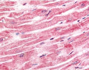
- Immunohistochemistry. Anti-Sprouty-4 antibody was used at a 2.5 μg/ml to detect cytoplasmic signal with moderate to strong staining and low background in a variety of tissues including heart, prostate, intestine, kidney and pancreas. Strong specific staining was noted at particular cell types in some tissues (i.e. Sertoli and Leydig cells). The antibody shows very good signal to background at 2.5 µg/ml. This image shows Sprouty-4 staining of human heart tissue. Tissue was formalin-fixed and paraffin embedded. Personal Communication, Tina Roush, LifeSpanBiosciences, Seattle, WA.
- Submitted by
- Acris Antibodies GmbH (provider)
- Main image

- Experimental details
- Immunohistochemistry. Anti-Sprouty-4 antibody shows strong cytoplasmic and membranous staining of tumor cells in human liver tissue. Tissue was formalin-fixed and paraffin embedded. Brown color indicates presence of protein, blue color shows cell nuclei. Personal Communication, Kenneth Wester, www.proteinatlas.org, Uppsala, Sweden.
