 Explore
 Validate
 Learn
Learn Western blot
Antibody data
- Antibody Data
- Antigen structure
- References [3]
- Comments [0]
- Validations
- Western blot [9]
- Immunocytochemistry [2]
- Immunohistochemistry [4]
Submit
Validation data
Reference
Comment
Report error
- Product number
- GTX101881 - Provider product page

- Provider
- GeneTex
- Proper citation
- GeneTex Cat#GTX101881, RRID:AB_1240876
- Product name
- GAD67 antibody
- Antibody type
- Polyclonal
- Reactivity
- Human, Mouse, Rat
- Host
- Rabbit
Submitted references Two Groups of eGFP-Expressing Neurons with Distinct Characteristics in the Neocortex of GIN Mice.
Age-related Changes in Auditory Cortex Without Detectable Peripheral Alterations: A Multi-level Study in Sprague-Dawley Rats.
Loss of phenotype of parvalbumin interneurons in rat prefrontal cortex is involved in antidepressant- and propsychotic-like behaviors following acute and repeated ketamine administration.
Wang C, Yu B, Li M, Zhao C, Roper SN, Chen H
Neuroscience 2019 Apr 15;404:268-281
Neuroscience 2019 Apr 15;404:268-281
Age-related Changes in Auditory Cortex Without Detectable Peripheral Alterations: A Multi-level Study in Sprague-Dawley Rats.
Occelli F, Hasselmann F, Bourien J, Eybalin M, Puel JL, Desvignes N, Wiszniowski B, Edeline JM, Gourévitch B
Neuroscience 2019 Apr 15;404:184-204
Neuroscience 2019 Apr 15;404:184-204
Loss of phenotype of parvalbumin interneurons in rat prefrontal cortex is involved in antidepressant- and propsychotic-like behaviors following acute and repeated ketamine administration.
Zhou Z, Zhang G, Li X, Liu X, Wang N, Qiu L, Liu W, Zuo Z, Yang J
Molecular neurobiology 2015 Apr;51(2):808-19
Molecular neurobiology 2015 Apr;51(2):808-19
No comments: Submit comment
Supportive validation
- Submitted by
- GeneTex (provider)
- Main image

- Experimental details
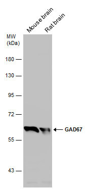
- GAD67 antibody detects GAD67 protein by Western blot analysis.A. 50 µg mouse brian lysate/extract7.5 % SDS-PAGEGAD67 antibody (GTX101881) dilution: 1:500
- Validation comment
- WB

- Submitted by
- GeneTex (provider)
- Main image

- Experimental details
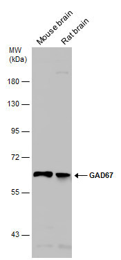
- GAD67 antibody detects GAD67 protein by Western blot analysis.A. 50 µg rat brain lysate/extract7.5 % SDS-PAGEGAD67 antibody (GTX101881) dilution: 1:500
- Validation comment
- WB

- Submitted by
- GeneTex (provider)
- Main image

- Experimental details
- GAD67 antibody detects GAD67 protein by Western blot analysis.A. 30 µg 293T whole cell lysate/extract B. 30 µg whole cell lysate/extract of human GAD1-transfected 293T cells7.5 % SDS-PAGEGAD67 antibody (GTX101881) dilution: 1:5000
- Validation comment
- WB

- Submitted by
- GeneTex (provider)
- Main image

- Experimental details
- GAD67 antibody detects GAD67 protein by Western blot analysis.A. 30 µg SK-N-SH whole cell lysate/extractB. 30 µg SK-N-AS whole cell lysate/extract7.5 % SDS-PAGEGAD67 antibody (GTX101881) dilution: 1:500
- Validation comment
- WB

- Submitted by
- GeneTex (provider)
- Main image

- Experimental details
- GAD67 antibody detects GAD67 protein by western blot analysis.A. 30 ?g 293T whole cell lysate/extractB. 30 ?g whole cell lysate/extract of human GAD1-transfected 293T cells7.5% SDS-PAGEGAD67 antibody (GTX101881) dilution: 1:5000The HRP-conjugated anti-rabbit IgG antibody (GTX213110-01) was used to detect the primary antibody.

- Submitted by
- GeneTex (provider)
- Main image

- Experimental details
- Various tissue extracts (50 ?g) were separated by 7.5% SDS-PAGE, and the membrane was blotted with GAD67 antibody (GTX101881) diluted at 1:1000. The HRP-conjugated anti-rabbit IgG antibody (GTX213110-01) was used to detect the primary antibody.
- Submitted by
- GeneTex (provider)
- Main image

- Experimental details
- GAD67 antibody detects GAD67 protein by western blot analysis.A. 30 ?g SK-N-SH whole cell lysate/extractB. 30 ?g SK-N-AS whole cell lysate/extract7.5% SDS-PAGEGAD67 antibody (GTX101881) dilution: 1:500The HRP-conjugated anti-rabbit IgG antibody (GTX213110-01) was used to detect the primary antibody.

- Submitted by
- GeneTex (provider)
- Main image

- Experimental details
- Whole cell extract (30 ?g) was separated by 7.5% SDS-PAGE, and the membrane was blotted with GAD67 antibody (GTX101881) diluted at 1:500.

- Submitted by
- GeneTex (provider)
- Main image

- Experimental details
- Various tissue extracts (50 ?g) were separated by 7.5% SDS-PAGE, and the membrane was blotted with GAD67 antibody (GTX101881) diluted at 1:1000.
Supportive validation
- Submitted by
- GeneTex (provider)
- Main image

- Experimental details
- GAD67 antibody detects GAD67 protein by immunofluorescent analysis. Sample: GAD67-transfected (right) or untransfected (left) 293T cells were fixed in 4% paraformaldehyde for 15 min. Green: GAD67 protein stained by GAD67 antibody (GTX101881) diluted at 1:500. Blue: Hoechst 33342 staining. Scale bar = 10 £gm.

- Submitted by
- GeneTex (provider)
- Main image

- Experimental details
- GAD67 antibody detects GAD67 protein at cytoplasm by immunofluorescent analysis.Sample: SK-N-SH cells were fixed in 4% paraformaldehyde at RT for 15 min.Green: GAD67 protein stained by GAD67 antibody (GTX101881) diluted at 1:200.Blue: Hoechst 33342 staining.

Supportive validation
- Submitted by
- GeneTex (provider)
- Main image

- Experimental details
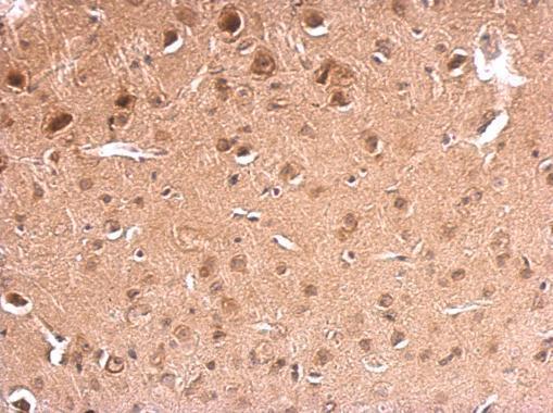
- GAD67 antibody detects GAD67 protein at on mouse fore brain by immunohistochemical analysis. Sample: Paraffin-embedded mouse fore brain. GAD67 antibody (GTX101881) dilution: 1:500.
- Submitted by
- GeneTex (provider)
- Main image

- Experimental details
- GAD67 antibody detects GAD67 protein expression by immunohistochemical analysis.Sample: Frozen sectioned E13.5 Rat brain. Green: GAD67 protein stained by GAD67 antibody (GTX101881) diluted at 1:250.Red: beta Tubulin 3/ TUJ1, a mature neuron marker, stained by beta Tubulin 3/ TUJ1 antibody [GT11710] (GTX631836) diluted at 1:500.Blue: Fluoroshield with DAPI (GTX30920).

- Submitted by
- GeneTex (provider)
- Main image

- Experimental details
- GAD67 antibody detects GAD67 protein at cytoplasm in rat brain by immunohistochemical analysis. Sample: Paraffin-embedded rat brain. GAD67 antibody (GTX101881) diluted at 1:500.

- Submitted by
- GeneTex (provider)
- Main image

- Experimental details
- GAD67 antibody detects GAD67 protein by immunohistochemical analysis.Sample: Frozen sectioned adult mouse retina. Green: GAD67 protein stained by GAD67 antibody (GTX101881) diluted at 1:250.Red: beta Tubulin 3/ TUJ1, stained by beta Tubulin 3/ TUJ1 antibody [GT11710] (GTX631836) diluted at 1:250.Blue: Fluoroshield with DAPI (GTX30920).
