 Explore
 Validate
 Learn
Learn Western blot
Antibody data
- Antibody Data
- Antigen structure
- References [3]
- Comments [0]
- Validations
- Western blot [2]
- Immunohistochemistry [1]
Submit
Validation data
Reference
Comment
Report error
- Product number
- AF2086 - Provider product page

- Provider
- R&D Systems
- Product name
- Human/Mouse/Rat GAD1/GAD67 Antibody
- Antibody type
- Polyclonal
- Description
- Antigen Affinity-purified. Detects human GAD1/GAD67 in direct ELISAs and detects human, mouse, and rat GAD1/GAD67 in Western blots. In Western blots, less than 1% cross-reactivity with recombinant human GAD2 is observed.
- Reactivity
- Human, Mouse, Rat
- Host
- Goat
- Conjugate
- Unconjugated
- Antigen sequence
Q99259- Isotype
- IgG
- Vial size
- 100 ug
- Concentration
- LYOPH
- Storage
- Use a manual defrost freezer and avoid repeated freeze-thaw cycles. 12 months from date of receipt, -20 to -70 °C as supplied. 1 month, 2 to 8 °C under sterile conditions after reconstitution. 6 months, -20 to -70 °C under sterile conditions after reconstitution.
Submitted references Alterations in a Unique Class of Cortical Chandelier Cell Axon Cartridges in Schizophrenia.
Transporters MCT8 and OATP1C1 maintain murine brain thyroid hormone homeostasis.
Locomotor deficiencies and aberrant development of subtype-specific GABAergic interneurons caused by an unliganded thyroid hormone receptor alpha1.
Rocco BR, DeDionisio AM, Lewis DA, Fish KN
Biological psychiatry 2017 Jul 1;82(1):40-48
Biological psychiatry 2017 Jul 1;82(1):40-48
Transporters MCT8 and OATP1C1 maintain murine brain thyroid hormone homeostasis.
Mayerl S, Müller J, Bauer R, Richert S, Kassmann CM, Darras VM, Buder K, Boelen A, Visser TJ, Heuer H
The Journal of clinical investigation 2014 May;124(5):1987-99
The Journal of clinical investigation 2014 May;124(5):1987-99
Locomotor deficiencies and aberrant development of subtype-specific GABAergic interneurons caused by an unliganded thyroid hormone receptor alpha1.
Wallis K, Sjögren M, van Hogerlinden M, Silberberg G, Fisahn A, Nordström K, Larsson L, Westerblad H, Morreale de Escobar G, Shupliakov O, Vennström B
The Journal of neuroscience : the official journal of the Society for Neuroscience 2008 Feb 20;28(8):1904-15
The Journal of neuroscience : the official journal of the Society for Neuroscience 2008 Feb 20;28(8):1904-15
No comments: Submit comment
Supportive validation
- Submitted by
- R&D Systems (provider)
- Main image
- Experimental details
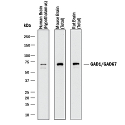
- Detection of Human, Mouse, and Rat GAD1/GAD67 by Western Blot. Western blot shows lysates of human brain (hypothalamus) tissue, mouse brain (total) tissue, and rat brain (total) tissue. PVDF membrane was probed with 1 µg/mL of Goat Anti-Human/Mouse/Rat GAD1/GAD67 Antigen Affinity-purified Polyclonal Antibody (Catalog # AF2086) followed by HRP-conjugated Anti-Goat IgG Secondary Antibody (Catalog # HAF017). A specific band was detected for GAD1/GAD67 at approximately 67 kDa (as indicated). This experiment was conducted under reducing conditions and using Immunoblot Buffer Group 1.

- Submitted by
- R&D Systems (provider)
- Main image
- Experimental details
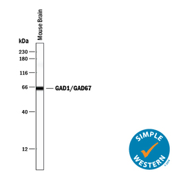
- Detection of Mouse GAD1/GAD67 by Simple WesternTM. Simple Western lane view shows lysates of mouse brain tissue, loaded at 0.2 mg/mL. A specific band was detected for GAD1/GAD67 at approximately 65 kDa (as indicated) using 1 µg/mL of Goat Anti-Human/Mouse/Rat GAD1/GAD67 Antigen Affinity-purified Polyclonal Antibody (Catalog # AF2086) followed by 1:50 dilution of HRP-conjugated Anti-Goat IgG Secondary Antibody (Catalog # HAF109). This experiment was conducted under reducing conditions and using the 12-230 kDa separation system.

Supportive validation
- Submitted by
- R&D Systems (provider)
- Main image
- Experimental details
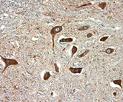
- GAD1/GAD67 in Human Spinal Cord. GAD1/GAD67 was detected in immersion fixed paraffin-embedded sections of human spinal cord using 15 µg/mL Goat Anti-Human/Mouse/Rat GAD1/GAD67 Antigen Affinity-purified Polyclonal Antibody (Catalog # AF2086) overnight at 4 °C. Tissue was stained with the Anti-Goat HRP-DAB Cell & Tissue Staining Kit (brown; Catalog # CTS008) and counterstained with hematoxylin (blue). View our protocol for Chromogenic IHC Staining of Paraffin-embedded Tissue Sections.
