Explore
 Validate
 Learn
Learn Western blot
 Immunocytochemistry
 Immunoprecipitation
Antibody data
- Antibody Data
- Antigen structure
- References [2]
- Comments [0]
- Validations
- Western blot [4]
- Immunocytochemistry [1]
- Immunohistochemistry [1]
Submit
Validation data
Reference
Comment
Report error
- Product number
- GTX105255 - Provider product page

- Provider
- GeneTex
- Proper citation
- GeneTex Cat#GTX105255, RRID:AB_1951346
- Product name
- PPP1A antibody
- Antibody type
- Polyclonal
- Reactivity
- Human, Rat
- Host
- Rabbit
Submitted references Carfilzomib induces leukaemia cell apoptosis via inhibiting ELK1/KIAA1524 (Elk-1/CIP2A) and activating PP2A not related to proteasome inhibition.
Phosphorylation of the Ndc80 complex protein, HEC1, by Nek2 kinase modulates chromosome alignment and signaling of the spindle assembly checkpoint.
Liu CY, Hsieh FS, Chu PY, Tsai WC, Huang CT, Yu YB, Huang TT, Ko PS, Hung MH, Wang WL, Shiau CW, Chen KF
British journal of haematology 2017 Jun;177(5):726-740
British journal of haematology 2017 Jun;177(5):726-740
Phosphorylation of the Ndc80 complex protein, HEC1, by Nek2 kinase modulates chromosome alignment and signaling of the spindle assembly checkpoint.
Wei R, Ngo B, Wu G, Lee WH
Molecular biology of the cell 2011 Oct;22(19):3584-94
Molecular biology of the cell 2011 Oct;22(19):3584-94
No comments: Submit comment
Enhanced validation
Supportive validation
- Submitted by
- GeneTex (provider)
- Enhanced method
- Genetic validation
- Main image
- Experimental details
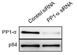
- WB to detect PP1 alpha from U2OS cells treated with control siRNA or PP1 alpha siRNA, using GTX105255 at 1:1000 dilution. Nuclear matrix protein p84 is a loading control, blotted with p84 antibody (clone 5E10) GTX70220 at 1:5000 dilution. The HRP-conjugated anti-rabbit IgG antibody (GTX213110-01) was used to detect the primary antibody.

Supportive validation
- Submitted by
- GeneTex (provider)
- Main image
- Experimental details
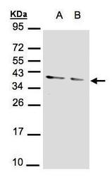
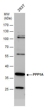
- Sample(30 ?g of whole cell lysate)A:293TB:A431(GTX27909)12% SDS PAGEGTX105255 diluted at 1:1000
- Validation comment
- WB
- Submitted by
- GeneTex (provider)
- Main image
- Experimental details
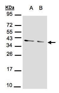
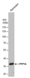
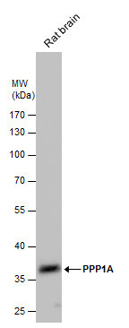
- PPP1A antibody detects PPP1A protein by western blot analysis. Rat tissue extracts (50 ?g) was separated by 10% SDS-PAGE, and the membrane was blotted with PPP1A antibody (GTX105255) diluted by 1:1000. The HRP-conjugated anti-rabbit IgG antibody (GTX213110-01) was used to detect the primary antibody.
- Submitted by
- GeneTex (provider)
- Main image
- Experimental details
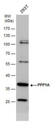
- PPP1A antibody detects PPP1A protein by western blot analysis. Whole cell extracts (30 ?g) was separated by 10% SDS-PAGE, and the membrane was blotted with PPP1A antibody (GTX105255) diluted by 1:1000. The HRP-conjugated anti-rabbit IgG antibody (GTX213110-01) was used to detect the primary antibody.
Supportive validation
- Submitted by
- GeneTex (provider)
- Main image

- Experimental details
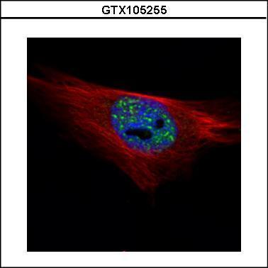
- Confocal immunofluorescence analysis (Olympus FV10i) of paraformaldehyde-fixed HeLa, using PPP1A(GTX105255) antibody (Green) at 1:500 dilution. Alpha-tubulin filaments were labeled with GTX11304 (Red) at 1:2500.
Supportive validation
- Submitted by
- GeneTex (provider)
- Main image
- Experimental details
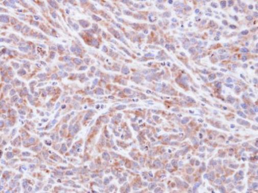
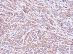
- Immunohistochemical analysis of paraffin-embedded SAS xenograft, using PPP1A(GTX105255) antibody at 1:100 dilution.