 Explore
 Validate
 Learn
Learn Western blot
Antibody data
- Antibody Data
- Antigen structure
- References [7]
- Comments [0]
- Validations
- Western blot [1]
- Immunocytochemistry [4]
- Immunohistochemistry [1]
- Other assay [1]
Submit
Validation data
Reference
Comment
Report error
- Product number
- MA5-12242 - Provider product page

- Provider
- Invitrogen Antibodies
- Product name
- 14-3-3 Pan Monoclonal Antibody (CG15)
- Antibody type
- Monoclonal
- Antigen
- Recombinant full-length protein
- Description
- MA5-12242 targets 14.3.3 pan in IP, ICC/IF, IHC (P) and WB applications and shows reactivity with Human and Mouse samples.
- Antibody clone number
- CG15
- Concentration
- 0.2 mg/mL
Submitted references p53/TAp63 and AKT regulate mammalian target of rapamycin complex 1 (mTORC1) signaling through two independent parallel pathways in the presence of DNA damage.
REDD1 deletion prevents dexamethasone-induced skeletal muscle atrophy.
mTOR activity under hypoxia.
Rescue of atypical protein kinase C in epithelia by the cytoskeleton and Hsp70 family chaperones.
The central proline rich region of POB1/REPS2 plays a regulatory role in epidermal growth factor receptor endocytosis by binding to 14-3-3 and SH3 domain-containing proteins.
Hypoxia regulates TSC1/2-mTOR signaling and tumor suppression through REDD1-mediated 14-3-3 shuttling.
Mechanism of Akt1 inhibition of breast cancer cell invasion reveals a protumorigenic role for TSC2.
Cam M, Bid HK, Xiao L, Zambetti GP, Houghton PJ, Cam H
The Journal of biological chemistry 2014 Feb 14;289(7):4083-94
The Journal of biological chemistry 2014 Feb 14;289(7):4083-94
REDD1 deletion prevents dexamethasone-induced skeletal muscle atrophy.
Britto FA, Begue G, Rossano B, Docquier A, Vernus B, Sar C, Ferry A, Bonnieu A, Ollendorff V, Favier FB
American journal of physiology. Endocrinology and metabolism 2014 Dec 1;307(11):E983-93
American journal of physiology. Endocrinology and metabolism 2014 Dec 1;307(11):E983-93
mTOR activity under hypoxia.
Vadysirisack DD, Ellisen LW
Methods in molecular biology (Clifton, N.J.) 2012;821:45-58
Methods in molecular biology (Clifton, N.J.) 2012;821:45-58
Rescue of atypical protein kinase C in epithelia by the cytoskeleton and Hsp70 family chaperones.
Mashukova A, Oriolo AS, Wald FA, Casanova ML, Kröger C, Magin TM, Omary MB, Salas PJ
Journal of cell science 2009 Jul 15;122(Pt 14):2491-503
Journal of cell science 2009 Jul 15;122(Pt 14):2491-503
The central proline rich region of POB1/REPS2 plays a regulatory role in epidermal growth factor receptor endocytosis by binding to 14-3-3 and SH3 domain-containing proteins.
Tomassi L, Costantini A, Corallino S, Santonico E, Carducci M, Cesareni G, Castagnoli L
BMC biochemistry 2008 Jul 22;9:21
BMC biochemistry 2008 Jul 22;9:21
Hypoxia regulates TSC1/2-mTOR signaling and tumor suppression through REDD1-mediated 14-3-3 shuttling.
DeYoung MP, Horak P, Sofer A, Sgroi D, Ellisen LW
Genes & development 2008 Jan 15;22(2):239-51
Genes & development 2008 Jan 15;22(2):239-51
Mechanism of Akt1 inhibition of breast cancer cell invasion reveals a protumorigenic role for TSC2.
Liu H, Radisky DC, Nelson CM, Zhang H, Fata JE, Roth RA, Bissell MJ
Proceedings of the National Academy of Sciences of the United States of America 2006 Mar 14;103(11):4134-9
Proceedings of the National Academy of Sciences of the United States of America 2006 Mar 14;103(11):4134-9
No comments: Submit comment
Supportive validation
- Submitted by
- Invitrogen Antibodies (provider)
- Main image

- Experimental details
- Western blot analysis was performed on whole cell extracts (30 µg lysate) of A-431 (Lane 1) A-375 (Lane 2), U-87 MG (Lane 3), MCF7 (Lane 4), HeLa (Lane 5) and HCT 116 (Lane 6). The blot was probed with anti-14-3-3 Pan Mouse Monoclonal Antibody (Product # MA5-12242, 1 µg/mL) and detected by chemiluminescence using Goat anti-Mouse IgG (H+L) Superclonal™ Secondary Antibody, HRP conjugate (Product # A28177, 0.25 µg/mL, 1:4000 dilution). A 28 kDa band corresponding to 14-3-3 Pan was detected across the cell lines tested.

Supportive validation
- Submitted by
- Invitrogen Antibodies (provider)
- Main image

- Experimental details
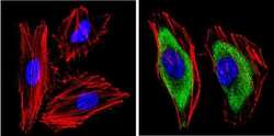
- Immunofluorescent analysis of 14.3.3 pan (green) showing staining in the cytoplasm of A431 cells. Formalin-fixed cells were permeabilized with 0.1% Triton X-100 in TBS for 5-10 minutes and blocked with 3% BSA-PBS for 30 minutes at room temperature. Cells were probed with a 14.3.3 pan monoclonal antibody (Product # MA5-12242) in 3% BSA-PBS at a dilution of 1:100 and incubated overnight at 4 ºC in a humidified chamber. Cells were washed with PBST and incubated with a DyLight-conjugated secondary antibody in PBS at room temperature in the dark. F-actin (red) was stained with a fluorescent red phalloidin and nuclei (blue) were stained with Hoechst or DAPI. Images were taken at a magnification of 60x.

- Submitted by
- Invitrogen Antibodies (provider)
- Main image
- Experimental details
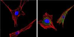
- Immunofluorescent analysis of 14.3.3 pan (green) showing staining in the cytoplasm of HeLa cells. Formalin-fixed cells were permeabilized with 0.1% Triton X-100 in TBS for 5-10 minutes and blocked with 3% BSA-PBS for 30 minutes at room temperature. Cells were probed with a 14.3.3 pan monoclonal antibody (Product # MA5-12242) in 3% BSA-PBS at a dilution of 1:100 and incubated overnight at 4 ºC in a humidified chamber. Cells were washed with PBST and incubated with a DyLight-conjugated secondary antibody in PBS at room temperature in the dark. F-actin (red) was stained with a fluorescent red phalloidin and nuclei (blue) were stained with Hoechst or DAPI. Images were taken at a magnification of 60x.

- Submitted by
- Invitrogen Antibodies (provider)
- Main image
- Experimental details
- Immunofluorescent analysis of 14.3.3 pan (green) showing staining in the cytoplasm of NIH-3T3 cells. Formalin-fixed cells were permeabilized with 0.1% Triton X-100 in TBS for 5-10 minutes and blocked with 3% BSA-PBS for 30 minutes at room temperature. Cells were probed with a 14.3.3 pan monoclonal antibody (Product # MA5-12242) in 3% BSA-PBS at a dilution of 1:100 and incubated overnight at 4 ºC in a humidified chamber. Cells were washed with PBST and incubated with a DyLight-conjugated secondary antibody in PBS at room temperature in the dark. F-actin (red) was stained with a fluorescent red phalloidin and nuclei (blue) were stained with Hoechst or DAPI. Images were taken at a magnification of 60x.

- Submitted by
- Invitrogen Antibodies (provider)
- Main image
- Experimental details
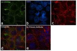
- Immunofluorescence analysis of 14-3-3 Pan was performed using 70% confluent log phase A-431 cells. The cells were fixed with 4% paraformaldehyde for 10 minutes, permeabilized with 0.1% Triton™ X-100 for 10 minutes, and blocked with 1% BSA for 1 hour at room temperature. The cells were labeled with 14-3-3 Mouse Monoclonal Antibody (Product # MA5-12242) at 5 µg/mL in 0.1% BSA and incubated overnight at 4 degree and then labeled with Goat anti-Mouse IgG (H+L) Superclonal™ Secondary Antibody, Alexa Fluor® 488 conjugate (Product # A28175) at a dilution of 1:2000 for 45 minutes at room temperature (Panel a: green). Nuclei (Panel b: blue) were stained with SlowFade® Gold Antifade Mountant with DAPI (Product # S36938). F-actin (Panel c: red) was stained with Rhodamine Phalloidin (Product # R415, 1:300). Panel d represents the merged image showing cytoplasmic localization. Panel e represents control cells with no primary antibody to assess background. The images were captured at 60X magnification.

Supportive validation
- Submitted by
- Invitrogen Antibodies (provider)
- Main image

- Experimental details
- Immunohistochemistry analysis of 14.3.3 pan showing positive staining in the cytoplasm of paraffin-treated Human colon carcinoma (right) compared with a negative control in the absence of primary antibody (left). To expose target proteins, antigen retrieval method was performed using 10mM sodium citrate (pH 6.0) microwaved for 8-15 min. Following antigen retrieval, tissues were blocked in 3% H2O2-methanol for 15 min at room temperature, washed with ddH2O and PBS, and then probed with a 14.3.3 pan monoclonal antibody (Product # MA5-12242) diluted by 3% BSA-PBS at a dilution of 1:20 overnight at 4°C in a humidified chamber. Tissues were washed extensively PBST and detection was performed using an HRP-conjugated secondary antibody followed by colorimetric detection using a DAB kit. Tissues were counterstained with hematoxylin and dehydrated with ethanol and xylene to prep for mounting.

Supportive validation
- Submitted by
- Invitrogen Antibodies (provider)
- Main image

- Experimental details
- Immunoprecipitation of 14.3.3 pan using 14.3.3 pan Monoclonal Antibody (Product # MA5-12242) on denatured Human HT29 Cells.
