 Explore
 Validate
 Learn
Learn Western blot
Antibody data
- Antibody Data
- Antigen structure
- References [2]
- Comments [0]
- Validations
- Western blot [3]
- Other assay [1]
Submit
Validation data
Reference
Comment
Report error
- Product number
- PA5-17288 - Provider product page

- Provider
- Invitrogen Antibodies
- Product name
- Arp3 Polyclonal Antibody
- Antibody type
- Polyclonal
- Antigen
- Synthetic peptide
- Description
- It is not recommended to aliquot this antibody. This antibody is not cross-reactive with endogenous levels of total ARP2 protein.
- Reactivity
- Human, Mouse, Rat
- Host
- Rabbit
- Isotype
- IgG
- Vial size
- 100 µL
- Concentration
- 66 µg/mL
- Storage
- -20°C
Submitted references The centrosome is an actin-organizing centre.
Enterohemorrhagic E. coli requires N-WASP for efficient type III translocation but not for EspFU-mediated actin pedestal formation.
Farina F, Gaillard J, Guérin C, Couté Y, Sillibourne J, Blanchoin L, Théry M
Nature cell biology 2016 Jan;18(1):65-75
Nature cell biology 2016 Jan;18(1):65-75
Enterohemorrhagic E. coli requires N-WASP for efficient type III translocation but not for EspFU-mediated actin pedestal formation.
Vingadassalom D, Campellone KG, Brady MJ, Skehan B, Battle SE, Robbins D, Kapoor A, Hecht G, Snapper SB, Leong JM
PLoS pathogens 2010 Aug 19;6(8):e1001056
PLoS pathogens 2010 Aug 19;6(8):e1001056
No comments: Submit comment
Supportive validation
- Submitted by
- Invitrogen Antibodies (provider)
- Main image
- Experimental details
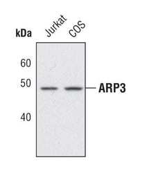
- Western blot analysis of ARP3 in extracts from Jurkat and COS cells using ARP3 polyclonal antibody (Product # PA5-17288).

- Submitted by
- Invitrogen Antibodies (provider)
- Main image
- Experimental details
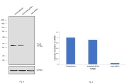
- Knockdown of Arp3 was achieved by transfecting MCF-7 cells with Arp3 specific siRNAs (Silencer® select Product # s19640, s19641). Western blot analysis (Fig. a) was performed using whole cell extracts from the MCF-7 knockdown cells (lane 3), non-specific scrambled siRNA transfected cells (lane 2) and untransfected cells (lane 1). The blots were probed with Arp3 Rabbit Polyclonal Antibody (Product # PA5-17288, 1:1000 dilution) and Goat anti-Rabbit IgG (H+L), Superclonal™ Recombinant Secondary Antibody, HRP (Product # A27036, 1:4000 dilution). Densitometric analysis of this western blot is shown in histogram (Fig. b). Decrease in signal upon siRNA mediated knock down confirms that antibody is specific to Arp3.

- Submitted by
- Invitrogen Antibodies (provider)
- Main image
- Experimental details
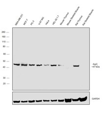
- Western blot was performed using Anti-Arp3 Polyclonal Antibody (Product # PA5-17288) and an ~47 kDa band was observed across the panel tested except Mouse and Rat Skeletal Muscle. Whole cell extracts (30 µg lysate) of MDA-MB-231 (Lane 1), MCF-7 (Lane 2), PC-3 (Lane 3), U-87 MG (Lane 4), HeLa (Lane 5), HEL 92.1.7 (Lane 6), tissue extracts (30 µg lysate) of Mouse Thymus (Lane 7), Mouse Skeletal Muscle (Lane 8), Rat Thymus (Lane 9) and Rat Skeletal Muscle (Lane 10) were electrophoresed using NuPAGE™ 10% Bis-Tris Protein Gel (Product # NP0302BOX). Resolved proteins were then transferred onto a nitrocellulose membrane (Product # IB23001) by iBlot® 2 Dry Blotting System (Product # IB21001). The blot was probed with the primary antibody (1:1000 dilution) and detected by chemiluminescence with Goat anti-Rabbit IgG (H+L), Superclonal™ Recombinant Secondary Antibody, HRP (Product # A27036, 1:4000 dilution) using the iBright FL 1000 (Product # A32752). Chemiluminescent detection was performed using Novex® ECL Chemiluminescent Substrate Reagent Kit (Product # WP20005).

Supportive validation
- Submitted by
- Invitrogen Antibodies (provider)
- Main image

- Experimental details
- NULL
