 Explore
 Validate
 Learn
Learn Western blot
 Immunocytochemistry
Antibody data
- Antibody Data
- Antigen structure
- References [7]
- Comments [0]
- Validations
- Western blot [2]
- Immunohistochemistry [1]
Submit
Validation data
Reference
Comment
Report error
- Product number
- MAB5718 - Provider product page

- Provider
- Novus Biologicals
- Product name
- Mouse Monoclonal GAPDH Antibody
- Antibody type
- Monoclonal
- Description
- Protein A or G purified from hybridoma culture supernatant. Detects human, mouse and rat GAPDH/G3PDH in Western blots. In direct ELISAs, 100% cross-reactivity with recombinant mouse GAPDH/G3PDH and no cross-reactivity with recombinant human GAPDH-2 is observed.
- Reactivity
- Human, Mouse, Rat
- Host
- Mouse
- Conjugate
- Unconjugated
- Isotype
- IgG
- Vial size
- 100 ug
- Storage
- Use a manual defrost freezer and avoid repeated freeze-thaw cycles. 12 months from date of receipt, -20 to -70 degreesC as supplied. 1 month, 2 to 8 degreesC under sterile conditions after reconstitution. 6 months, -20 to -70 degreesC under sterile conditions after reconstitution.
Submitted references Geranylgeraniol (GGOH) as a Mevalonate Pathway Activator in the Rescue of Bone Cells Treated with Zoledronic Acid: An In Vitro Study.
Metformin exhibited anticancer activity by lowering cellular cholesterol content in breast cancer cells.
Leptin enhances cytokine/chemokine production by normal lung fibroblasts by binding to leptin receptor.
A modifier in the 129S2/SvPasCrl genome is responsible for the viability of Notch1[12f/12f] mice.
Characterization of the Giardia intestinalis secretome during interaction with human intestinal epithelial cells: The impact on host cells.
The transcription factor MITF is a critical regulator of GPNMB expression in dendritic cells.
Normal leptin expression, lower adipogenic ability, decreased leptin receptor and hyposensitivity to Leptin in Adolescent Idiopathic Scoliosis.
Fliefel RM, Entekhabi SA, Ehrenfeld M, Otto S
Stem cells international 2019;2019:4351327
Stem cells international 2019;2019:4351327
Metformin exhibited anticancer activity by lowering cellular cholesterol content in breast cancer cells.
Sharma A, Bandyopadhayaya S, Chowdhury K, Sharma T, Maheshwari R, Das A, Chakrabarti G, Kumar V, Mandal CC
PloS one 2019;14(1):e0209435
PloS one 2019;14(1):e0209435
Leptin enhances cytokine/chemokine production by normal lung fibroblasts by binding to leptin receptor.
Watanabe K, Suzukawa M, Arakawa S, Kobayashi K, Igarashi S, Tashimo H, Nagai H, Tohma S, Nagase T, Ohta K
Allergology international : official journal of the Japanese Society of Allergology 2019 Sep;68S:S3-S8
Allergology international : official journal of the Japanese Society of Allergology 2019 Sep;68S:S3-S8
A modifier in the 129S2/SvPasCrl genome is responsible for the viability of Notch1[12f/12f] mice.
Varshney S, Wei HX, Batista F, Nauman M, Sundaram S, Siminovitch K, Tanwar A, Stanley P
BMC developmental biology 2019 Oct 7;19(1):19
BMC developmental biology 2019 Oct 7;19(1):19
Characterization of the Giardia intestinalis secretome during interaction with human intestinal epithelial cells: The impact on host cells.
Ma'ayeh SY, Liu J, Peirasmaki D, Hörnaeus K, Bergström Lind S, Grabherr M, Bergquist J, Svärd SG
PLoS neglected tropical diseases 2017 Dec;11(12):e0006120
PLoS neglected tropical diseases 2017 Dec;11(12):e0006120
The transcription factor MITF is a critical regulator of GPNMB expression in dendritic cells.
Gutknecht M, Geiger J, Joas S, Dörfel D, Salih HR, Müller MR, Grünebach F, Rittig SM
Cell communication and signaling : CCS 2015 Mar 24;13:19
Cell communication and signaling : CCS 2015 Mar 24;13:19
Normal leptin expression, lower adipogenic ability, decreased leptin receptor and hyposensitivity to Leptin in Adolescent Idiopathic Scoliosis.
Liang G, Gao W, Liang A, Ye W, Peng Y, Zhang L, Sharma S, Su P, Huang D
PloS one 2012;7(5):e36648
PloS one 2012;7(5):e36648
No comments: Submit comment
Supportive validation
- Submitted by
- Novus Biologicals (provider)
- Main image
- Experimental details
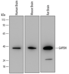
- Detection of Human, Mouse, and Rat GAPDH/G3PDH by Western Blot. Western blot shows lysates of human brain tissue, mouse brain tissue, and rat brain tissue. PVDF Membrane was probed with 0.05 µg/mL of Mouse Anti-Human/Mouse/Rat GAPDH/G3PDH Monoclonal Antibody (Catalog # MAB5718) followed by HRP-conjugated Anti-Mouse IgG Secondary Antibody (Catalog # HAF007). A specific band was detected for GAPDH/G3PDH at approximately 39 kDa (as indicated). This experiment was conducted under reducing conditions and using Immunoblot Buffer Group 2.

- Submitted by
- Novus Biologicals (provider)
- Main image
- Experimental details
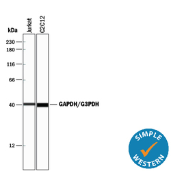
- Detection of Human and Mouse GAPDH/G3PDH by Simple WesternTM. Simple Western lane view shows lysates of Jurkat human acute T cell leukemia cell line and C2C12 mouse myoblast cell line, loaded at 0.2 mg/mL. A specific band was detected for GAPDH/G3PDH at approximately 41 kDa (as indicated) using 0.25 µg/mL of Mouse Anti-Human/Mouse/Rat GAPDH/G3PDH Monoclonal Antibody (Catalog # MAB5718). This experiment was conducted under reducing conditions and using the 12-230 kDa separation system.

Supportive validation
- Submitted by
- Novus Biologicals (provider)
- Main image

- Experimental details
- GAPDH/G3PDH in Human Kidney. GAPDH/G3PDH was detected in immersion fixed paraffin-embedded sections of human kidney using Mouse Anti-Human/Mouse/Rat GAPDH/G3PDH Monoclonal Antibody (Catalog # MAB5718) at 15 µg/mL overnight at 4 °C. Before incubation with the primary antibody, tissue was subjected to heat-induced epitope retrieval using Antigen Retrieval Reagent-Basic (Catalog # CTS013). Tissue was stained using the Anti-Mouse HRP-DAB Cell & Tissue Staining Kit (brown; Catalog # CTS002) and counterstained with hematoxylin (blue). Specific staining was localized to cytoplasm and nuclei. View our protocol for Chromogenic IHC Staining of Paraffin-embedded Tissue Sections.
