Explore
 Validate
 Learn
LearnMA5-14739
antibody from Invitrogen Antibodies
Targeting: TBP
GTF2D1, SCA17, TFIID
 Western blot Immunocytochemistry
 Immunoprecipitation
 Immunohistochemistry Flow cytometry Chromatin Immunoprecipitation
Antibody data
- Antibody Data
- Antigen structure
- References [4]
- Comments [0]
- Validations
- Western blot [4]
- Immunocytochemistry [1]
Submit
Validation data
Reference
Comment
Report error
- Product number
- MA5-14739 - Provider product page

- Provider
- Invitrogen Antibodies
- Product name
- Anti-TBP Monoclonal Antibody (51841)
- Antibody type
- Monoclonal
- Antigen
- Synthetic peptide
- Description
- Heat mediated antigen retrieval recommended prior to tissue staining. This antibody is predicted to react with chicken, cow, Xenopus laevis, chimpanzee and zebrafish based on sequence homology.
- Reactivity
- Human, Mouse, Rat
- Host
- Mouse
- Isotype
- IgG
- Antibody clone number
- 51841
- Vial size
- 100 µg
- Concentration
- 1.0 mg/mL
- Storage
- -20°C or -80°C if preferred
Submitted references Pigment Epithelium-Derived Factor (PEDF) mediates cartilage matrix loss in an age-dependent manner under inflammatory conditions.
mTORC1 Down-Regulates Cyclin-Dependent Kinase 8 (CDK8) and Cyclin C (CycC).
Evaluation of the lipopolysaccharide-induced transcription of the human TREM-1 gene in vitamin D3-matured THP-1 macrophage-like cells.
ESRP2 controls an adult splicing programme in hepatocytes to support postnatal liver maturation.
Nakamura DS, Hollander JM, Uchimura T, Nielsen HC, Zeng L
BMC musculoskeletal disorders 2017 Jan 25;18(1):39
BMC musculoskeletal disorders 2017 Jan 25;18(1):39
mTORC1 Down-Regulates Cyclin-Dependent Kinase 8 (CDK8) and Cyclin C (CycC).
Feng D, Youn DY, Zhao X, Gao Y, Quinn WJ 3rd, Xiaoli AM, Sun Y, Birnbaum MJ, Pessin JE, Yang F
PloS one 2015;10(6):e0126240
PloS one 2015;10(6):e0126240
Evaluation of the lipopolysaccharide-induced transcription of the human TREM-1 gene in vitamin D3-matured THP-1 macrophage-like cells.
Hosoda H, Tamura H, Nagaoka I
International journal of molecular medicine 2015 Nov;36(5):1300-10
International journal of molecular medicine 2015 Nov;36(5):1300-10
ESRP2 controls an adult splicing programme in hepatocytes to support postnatal liver maturation.
Bhate A, Parker DJ, Bebee TW, Ahn J, Arif W, Rashan EH, Chorghade S, Chau A, Lee JH, Anakk S, Carstens RP, Xiao X, Kalsotra A
Nature communications 2015 Nov 4;6:8768
Nature communications 2015 Nov 4;6:8768
No comments: Submit comment
Supportive validation
- Submitted by
- Invitrogen Antibodies (provider)
- Main image
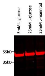
- Experimental details
- Western blot analysis of TATA binding protein (TBP) was performed by loading 25 µg of human retinal microvascular endothelial cell nuclear lysates, from cells treated with 5mM L-glucose (lane 1), 25mM L-glucose (lane 2), and 25mM L-mannitol (lane 3) per well onto an SDS-PAGE gel. Proteins were transferred to a nitrocellulose membrane and blocked for 1 hour at room temperature. The membrane was probed with a TBP monoclonal antibody (Product # MA5-14739) at a dilution of 1:1000 overnight at 4C, washed in PBST, and probed with a near-IR-conjugated donkey anti-mouse IgG secondary antibody at a dilution of 1:5000 for 1 hour at room temperature. Detection was performed using a near-IR imaging system. Data courtesy of the Innovators Program. NOTE: Nuclear lysates were generated using NE-PER Nuclear and Cytoplasmic Extraction Reagents (Product # 78833).

- Submitted by
- Invitrogen Antibodies (provider)
- Main image

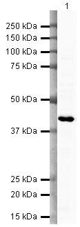
- Experimental details
- Western blot analysis of HeLa Whole Cell Lysate using Product # MA5-14739, TATA binding protein TBP primary antibody at a dilution of 1 µg/mL. Blot treated with a secondary HRP-conjugated Goat polyclonal anti-Mouse antibody was used at a dilution of 1:3000.
- Submitted by
- Invitrogen Antibodies (provider)
- Main image
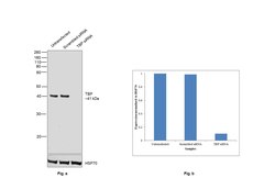
- Experimental details
- Knockdown of TBP was achieved by transfecting HeLa with TBP specific siRNAs (Silencer® select Product # s13826, s13827). Western blot analysis (Fig. a) was performed using nuclear enriched extracts from the TBP knockdown cells (lane 3), non-targeting scrambled siRNA transfected cells (lane 2) and untransfected cells (lane 1). The blot was probed with TBP Monoclonal Antibody (51841) (Product # MA5-14739, 1:1000 dilution) and Goat anti-Mouse IgG (H+L) Superclonal™ Recombinant Secondary Antibody, HRP (Product # A28177, 1:4000 dilution). Densitometric analysis of this western blot is shown in the histogram (Fig. b). The decrease in signal upon siRNA mediated knockdown confirms that the antibody is specific to TBP.

- Submitted by
- Invitrogen Antibodies (provider)
- Main image
- Experimental details
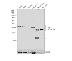
- Western blot was performed using Anti-TBP Monoclonal Antibody (51841) (Product # MA5-14739) and a 41kDa band corresponding to TBP was observed across all cell lines tested, except NIH/3T3 and mouse and rat testes tissues where a 37kDa band was observed. A ~25 kDa band (*) corresponding to IgG was also observed in the mouse testis sample. Nuclear enriched extracts (30 µg lysate) of K-562 (Lane 1), HeLa (Lane 2), NIH/3T3 (Lane 3), HEL 92.1.7 (Lane 4), Mouse Testis (Lane 5) and Rat Testis (Lane 6) were electrophoresed using NuPAGE™ 10% Bis-Tris Protein Gel (Product # NP0302BOX). Resolved proteins were then transferred onto a Nitrocellulose membrane (Product # IB23001) by iBlot® 2 Dry Blotting System (Product # IB21001). The blot was probed with the primary antibody (1:1000 dilution) and detected by chemiluminescence with Goat anti-Mouse IgG (H+L) Superclonal™ Recombinant Secondary Antibody, HRP (Product # A28177,1:4000 dilution) using the iBright FL 1000 (Product # A32752). Chemiluminescent detection was performed using SuperSignal™ West Dura Extended Duration Substrate (Product # 34076).

Supportive validation
- Submitted by
- Invitrogen Antibodies (provider)
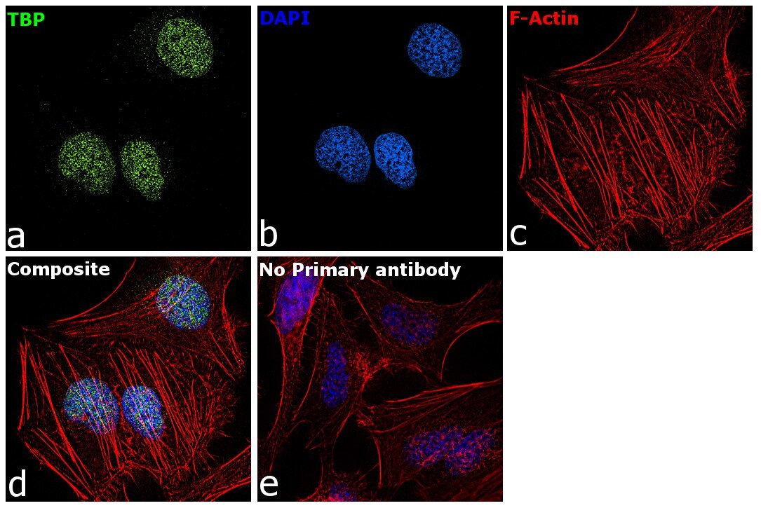
- Main image

- Experimental details
- Immunofluorescence analysis of TBP was performed using 70% confluent log phase HeLa cells. The cells were fixed with 4% paraformaldehyde for 10 minutes, permeabilized with 0.1% Triton™ X-100 for 15 minutes, and blocked with 2% BSA for 45 minutes at room temperature. The cells were labeled with TBP Monoclonal Antibody (51841) (Product # MA5-14739) at 1 µg/mL concentration in 0.1% BSA, incubated at 4 degree celsius overnight and then labeled with Donkey anti-Mouse IgG (H+L) Highly Cross-Adsorbed Secondary Antibody, Alexa Fluor Plus 488 (Product # A32766), (1:2000 dilution), for 45 minutes at room temperature (Panel a: Green). Nuclei (Panel b:Blue) were stained with ProLong™ Diamond Antifade Mountant with DAPI (Product # P36962). F-actin (Panel c: Red) was stained with Rhodamine Phalloidin (Product # R415, 1:300 dilution). Panel d represents the merged image showing nuclear localization. Panel e represents control cells with no primary antibody to assess background. The images were captured at 60X magnification.