Explore
 Validate
 Learn
Learn Western blot
Antibody data
- Antibody Data
- Antigen structure
- References [0]
- Comments [0]
- Validations
- Western blot [2]
- Immunocytochemistry [1]
- Immunohistochemistry [1]
- Flow cytometry [1]
Submit
Validation data
Reference
Comment
Report error
- Product number
- MA5-34787 - Provider product page

- Provider
- Invitrogen Antibodies
- Product name
- TYROBP Recombinant Rabbit Monoclonal Antibody (JG38-70)
- Antibody type
- Monoclonal
- Antigen
- Recombinant full-length protein
- Description
- Positive Control: THP-1, K562, human lung cancer tissue.
- Reactivity
- Human
- Host
- Rabbit
- Isotype
- IgG
- Antibody clone number
- JG38-70
- Vial size
- 100 µL
- Concentration
- 1 mg/mL
- Storage
- -20° C, Avoid Freeze/Thaw Cycles, store in dark
No comments: Submit comment
Supportive validation
- Submitted by
- Invitrogen Antibodies (provider)
- Main image

- Experimental details
- Western blot analysis of DAP12 in THP-1 cell. Samples were incubated with DAP12 monoclonal antibody (Product # MA5-34787), at a dilution of 1:500.

- Submitted by
- Invitrogen Antibodies (provider)
- Main image

- Experimental details
- Western blot was performed using TYROBP Recombinant Rabbit Monoclonal Antibody (JG38-70) (Product # MA5-34787) and a ~12 kDa band corresponding to Tyrobp was observed in THP1 and Higher expression was observed in THP1 differentiated to Macrophages except MCF7, HACAT and RAW 264.7 were reported to be negative. Whole cell Lysate (30 µg lysate) of THP-1 (Lane 1), U-937 (Lane 1), THP-1 (Lane 2), THP-1 (Lane 3), MCF7 (Lane 3), MCF7 (Lane 4), HaCaT (Lane 4), HaCaT (Lane 5), RAW 264.7 (Lane 5), RAW 264.7 (Lane 6) were electrophoresed using Novex™ 16% Tricine Protein Gel (Product # EC6695BOX), 10 well. Resolved proteins were then transferred onto a nitrocellulose membrane (Product # IB23001) by iBlot® 2 Dry Blotting System (Product # IB21001). The blot was probed with the primary antibody (1:1000 dilution) and detected by chemiluminescence with Goat anti-Rabbit IgG (H+L) Superclonal™ Recombinant Secondary Antibody, HRP (Product # A27036, 1:20,000 dilution) using the iBright™ FL1500 Imaging System (Product # A44115). Chemiluminescent detection was performed using SuperSignal™ West Atto Ultimate Sensitivity Substrate (Product # A38556).A single non specific band was observed (*).

Supportive validation
- Submitted by
- Invitrogen Antibodies (provider)
- Main image

- Experimental details
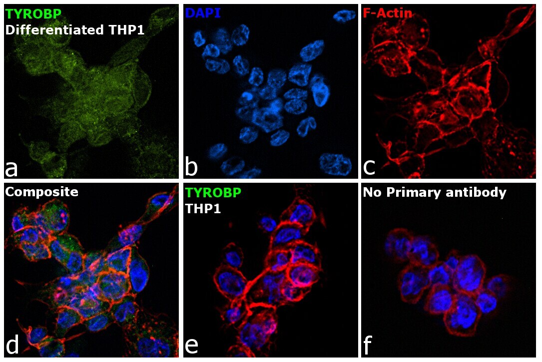
- Immunofluorescence analysis of TYROBP is performed using THP-1 cells and THP1 differentiated to Macrophages. The cells were fixed with 4% paraformaldehyde for 10 minutes, permeabilized with 0.1% Triton™ X-100 for 10 minutes, and blocked with 2% BSA for 45 minutes at room temperature. The cells were labeled with TYROBP Recombinant Rabbit Monoclonal Antibody (JG38-70) (Product # MA5-34787) at 1:100 dilution in 0.1% BSA, incubated at 4 degree celsius overnight and then labeled with Donkey anti-Rabbit IgG (H+L) Highly Cross-Adsorbed Secondary Antibody, Alexa Fluor™ Plus 488 (Product # A32790, 1:2000 dilution), for 45 minutes at room temperature (Panel a: Green). Nuclei (Panel b: Blue) were stained with ProLong™ Diamond Antifade Mountant with DAPI (Product # P36962). F-actin (Panel c: Red) was stained withAlexa Fluor™ 488 Phalloidin (Product # A12379, 1:300). Panel d represents the merged image showing Plasma Membrane localization. Panel e represents THP1 undifferentiated cells. Panel f represents control cells with no primary antibody to assess background. The images were captured at 60X magnification.
Supportive validation
- Submitted by
- Invitrogen Antibodies (provider)
- Main image
- Experimental details
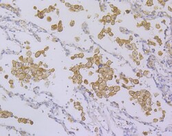
- Immunohistochemistry analysis of DAP12 in paraffin-embedded human lung cancer tissue. Samples were incubated with DAP12 monoclonal antibody (Product # MA5-34787), and followed by hematoxylin.

Supportive validation
- Submitted by
- Invitrogen Antibodies (provider)
- Main image
- Experimental details
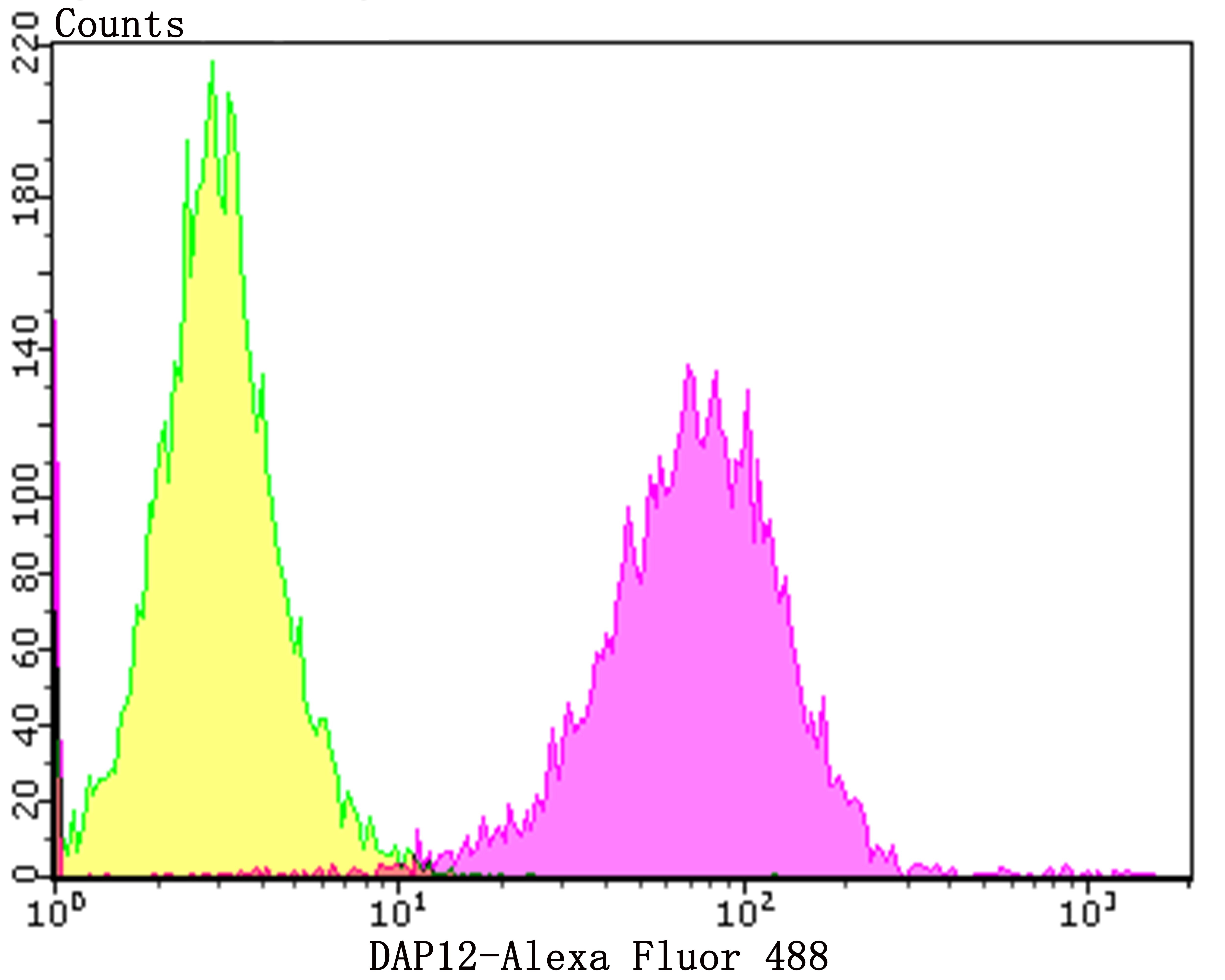
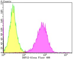
- Flow cytometry of DAP12 in K562 cells (purple) compared with an unlabelled control (cells without incubation with primary antibody; yellow). Samples were incubated with DAP12 monoclonal antibody (Product # MA5-34787) at a dilution of 1:100, followed by Alexa Fluor 488-conjugated goat anti-rabbit IgG.