Explore
 Validate
 Learn
Learn Western blot
 Immunoprecipitation
Antibody data
- Antibody Data
- Antigen structure
- References [2]
- Comments [0]
- Validations
- Western blot [1]
- Immunocytochemistry [1]
Submit
Validation data
Reference
Comment
Report error
- Product number
- AP08739PU-N - Provider product page

- Provider
- Acris Antibodies GmbH
- Proper citation
- Acris Antibodies GmbH Cat#AP08739PU-N, RRID:AB_1975708
- Product name
- anti Synapsin-1
- Antibody type
- Polyclonal
- Antigen
- Native protein purified from bovine brain.
- Reactivity
- Human, Mouse, Rat
- Host
- Rabbit
- Isotype
- IgG
- Vial size
- 10 µg
Submitted references Ca2+-independent activation of Ca2+/calmodulin-dependent protein kinase II bound to the C-terminal domain of CaV2.1 calcium channels.
Long term synaptic depression that is associated with GluR1 dephosphorylation but not alpha-amino-3-hydroxy-5-methyl-4-isoxazolepropionic acid (AMPA) receptor internalization.
Magupalli VG, Mochida S, Yan J, Jiang X, Westenbroek RE, Nairn AC, Scheuer T, Catterall WA
The Journal of biological chemistry 2013 Feb 15;288(7):4637-48
The Journal of biological chemistry 2013 Feb 15;288(7):4637-48
Long term synaptic depression that is associated with GluR1 dephosphorylation but not alpha-amino-3-hydroxy-5-methyl-4-isoxazolepropionic acid (AMPA) receptor internalization.
Davies KD, Goebel-Goody SM, Coultrap SJ, Browning MD
The Journal of biological chemistry 2008 Nov 28;283(48):33138-46
The Journal of biological chemistry 2008 Nov 28;283(48):33138-46
No comments: Submit comment
Supportive validation
- Submitted by
- Acris Antibodies GmbH (provider)
- Main image
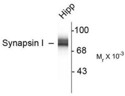
- Experimental details
- Figure 1. Western blot of 10 µg of Rat Hippocampal (Hipp) lysate showing specific immunolabeling of the ~78k Synapsin I doublet protein.

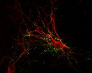
Supportive validation
- Submitted by
- Acris Antibodies GmbH (provider)
- Main image

- Experimental details
- Figure 2. Immunochemical Staining of Cultured Caudate Neurons with anti-Synapsin Antibody (Cat#AP08739PU-N) (Green) and anti-MAP antibody (Red).