Explore
 Validate
 Learn
Learn Western blot
Antibody data
- Antibody Data
- Antigen structure
- References [1]
- Comments [0]
- Validations
- Western blot [6]
- Immunocytochemistry [2]
Submit
Validation data
Reference
Comment
Report error
- Product number
- MA1-23328 - Provider product page

- Provider
- Invitrogen Antibodies
- Product name
- XPC Monoclonal Antibody (3.26)
- Antibody type
- Monoclonal
- Antigen
- Purifed from natural sources
- Description
- Recommended positive controls: HeLa whole cell extract.
- Antibody clone number
- 3.26
- Concentration
- 1.15 mg/mL
Submitted references CRISPR-Pass: Gene Rescue of Nonsense Mutations Using Adenine Base Editors.
Lee C, Hyun Jo D, Hwang GH, Yu J, Kim JH, Park SE, Kim JS, Kim JH, Bae S
Molecular therapy : the journal of the American Society of Gene Therapy 2019 Aug 7;27(8):1364-1371
Molecular therapy : the journal of the American Society of Gene Therapy 2019 Aug 7;27(8):1364-1371
No comments: Submit comment
Supportive validation
- Submitted by
- Invitrogen Antibodies (provider)
- Main image
- Experimental details
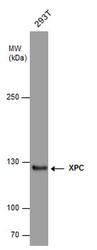
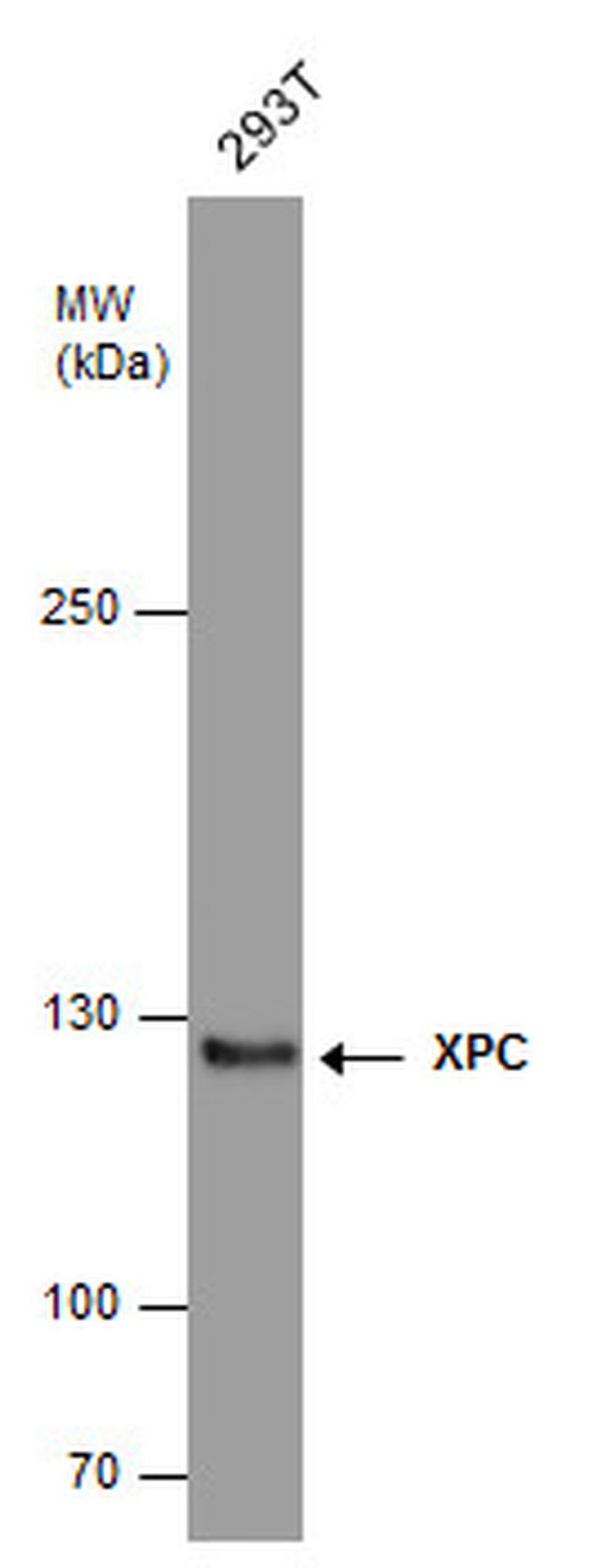
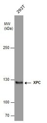
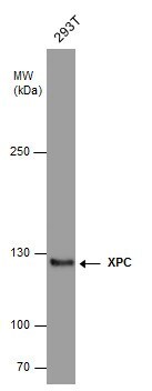
- Western blot analysis of XPC in whole cell extracts (30 µg). Samples was separated by 5% SDS-PAGE and the membrane was probed with XPC Monoclonal antibody (Product # MA1-23328) at a dilution of 1:500.
- Submitted by
- Invitrogen Antibodies (provider)
- Main image

- Experimental details
- Western blot of XPC in Raji whole cell extract using a XPC monoclonal antibody (Product # MA1-23328) at a dilution of 1:1000 (Lane 1) and 1:500 (Lane 2).

- Submitted by
- Invitrogen Antibodies (provider)
- Main image
- Experimental details
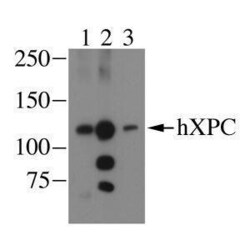
- Western blot image of human XPC from HeLa whole cell lysate detected using XPC Monoclonal Antibody (3.26) (Product # MA1-23328) (lane 3).

- Submitted by
- Invitrogen Antibodies (provider)
- Main image
- Experimental details
- XPC Monoclonal Antibody (3.26) detects XPC protein by western blot analysis. Whole cell extracts (30 µg) was separated by 5% SDS-PAGE, and the membrane was blotted with XPC Monoclonal Antibody (3.26) (Product # MA1-23328) diluted at 1:500. The HRP-conjugated anti-mouse IgG antibody was used to detect the primary antibody.
- Submitted by
- Invitrogen Antibodies (provider)
- Main image
- Experimental details
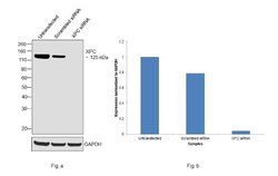
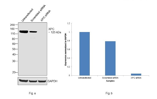
- Knockdown of XPC was achieved by transfecting HeLa with XPC specific siRNAs (Silencer® select Product # s14930, s533962). Western blot analysis (Fig. a) was performed using whole cell extracts from the XPC knockdown cells (Lane 3), non-specific scrambled siRNA transfected cells (Lane 2) and untransfected cells (Lane 1). The blot was probed with XPC Monoclonal Antibody (3.26) (Product # MA1-23328, 1:1000 dilution) and Goat anti-Mouse IgG (H+L), Superclonal™ Recombinant Secondary Antibody, HRP conjugate (Product # A28177, 0.25ug/ml, 1:4000 dilution). Densitometric analysis of this western blot is shown in histogram (Fig. b). Decrease in signal upon siRNA mediated knock down confirms that antibody is specific to XPC.
- Submitted by
- Invitrogen Antibodies (provider)
- Main image
- Experimental details
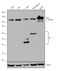
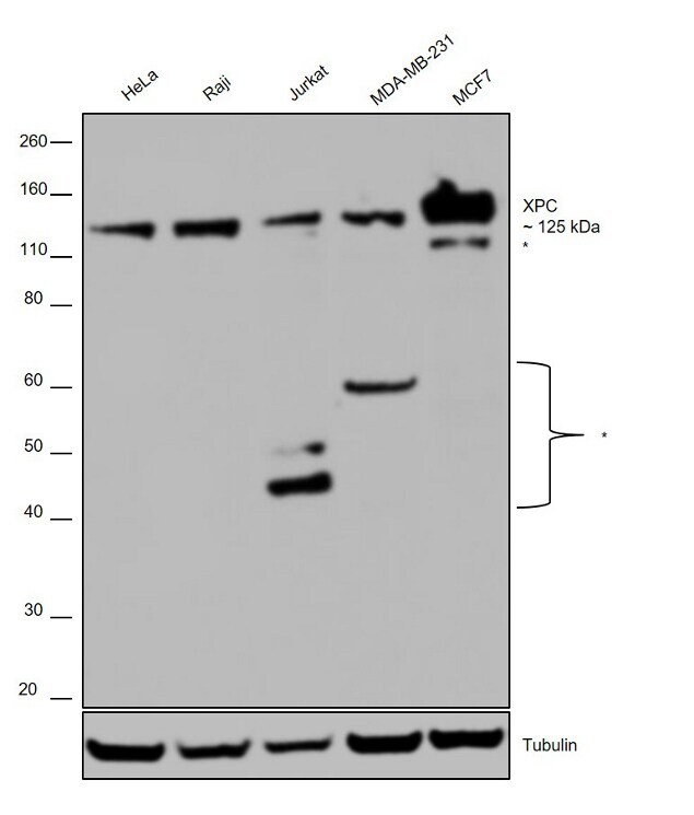
- Western blot was performed using Anti-XPC Monoclonal Antibody (Product # MA1-23328) and a ~125 kDa band corresponding to XPC was observed across cell lines along with certain uncharacterized bands (*) at different molecular weights. Membrane enriched extracts (30 µg lysate) of HeLa (Lane 1), Raji (Lane 2), Jurkat (Lane 3), MDA-MB-231 (Lane 4) and MCF7 (Lane 5) were electrophoresed using Novex® NuPAGE® 4-12 % Bis-Tris gel (Product # NP0322BOX). Resolved proteins were then transferred onto a nitrocellulose membrane (Product # IB23001) by iBlot® 2 Dry Blotting System (Product # IB21001). The blot was probed with the primary antibody (1:1000 dilution) and detected by chemiluminescence Goat anti-Mouse IgG (H+L), Superclonal™ Recombinant Secondary Antibody, HRP (Product # A28177), 1:4000 dilution) using the iBright FL 1000 (Product # A32752). Chemiluminescent detection was performed using Novex® ECL Chemiluminescent Substrate Reagent Kit (Product # WP20005).
Supportive validation
- Submitted by
- Invitrogen Antibodies (provider)
- Main image
- Experimental details
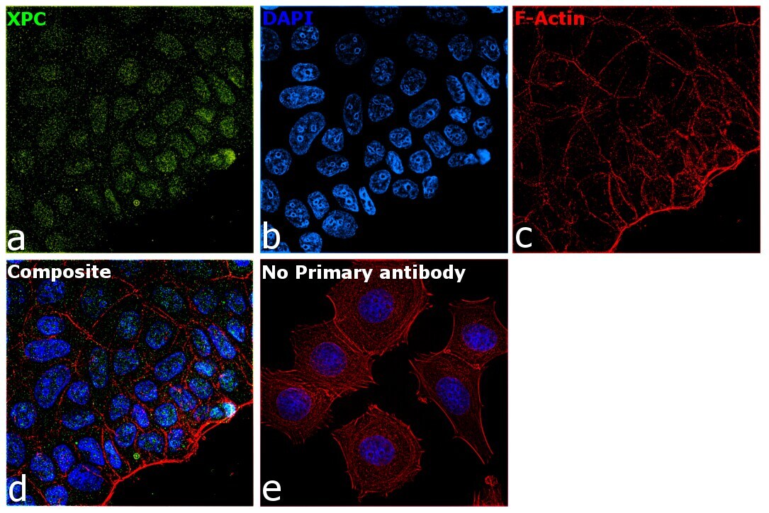
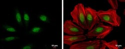
- XPC Monoclonal Antibody (3.26) detects XPC protein at nucleus by immunofluorescent analysis. Sample: HeLa cells were fixed in 4% paraformaldehyde at RT for 15 min. Green: XPC protein stained by XPC Monoclonal Antibody (3.26) (Product # MA1-23328) diluted at 1:100. Red: Phalloidin, a cytoskeleton marker, diluted at 1:200. Scale bar = 10 µm.

- Submitted by
- Invitrogen Antibodies (provider)
- Main image

- Experimental details
- Immunofluorescence analysis of XPC was performed using 70% confluent log phase MCF7 cells. The cells were fixed with 4% paraformaldehyde for 10 minutes, permeabilized with 0.1% Triton™ X-100 for 15 minutes, and blocked with 2% BSA for 1 hour at room temperature. The cells were labeled with XPC Monoclonal Antibody (3.26) (Product # MA1-23328) at 5 µg/mL in 0.1% BSA, incubated at 4 degree Celsius overnight and then with Goat anti-Mouse IgG (H+L), Superclonal™ Recombinant Secondary Antibody, Alexa Fluor 488 conjugate (Product # A28175) at a dilution of 1:2000 for 45 minutes at room temperature (Panel a: green). Nuclei (Panel b: blue) were stained with SlowFade® Gold Antifade Mountant with DAPI (Product # S36938). F-actin (Panel c: red) was stained with Rhodamine Phalloidin (Product # R415, 1:300). Panel d represents the merged image showing staining in nucleus. Panel e represents control cells with no primary antibody to assess background. The images were captured at 60X magnification.