 Explore
 Validate
 Learn
Learn Western blot
Antibody data
- Antibody Data
- Antigen structure
- References [1]
- Comments [0]
- Validations
- Western blot [6]
- Immunocytochemistry [4]
- Immunohistochemistry [1]
- Other assay [1]
Submit
Validation data
Reference
Comment
Report error
- Product number
- PA5-22127 - Provider product page

- Provider
- Invitrogen Antibodies
- Product name
- RAB6A Polyclonal Antibody
- Antibody type
- Polyclonal
- Antigen
- Recombinant full-length protein
- Description
- Recommended positive controls: 293T, A549, H1299, HCT116, mouse brain, rat brain.
- Concentration
- 0.21 mg/mL
Submitted references Exocytosis of Progeny Infectious Varicella-Zoster Virus Particles via a Mannose-6-Phosphate Receptor Pathway without Xenophagy following Secondary Envelopment.
Girsch JH, Jackson W, Carpenter JE, Moninger TO, Jarosinski KW, Grose C
Journal of virology 2020 Jul 30;94(16)
Journal of virology 2020 Jul 30;94(16)
No comments: Submit comment
Supportive validation
- Submitted by
- Invitrogen Antibodies (provider)
- Main image
- Experimental details
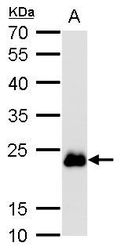
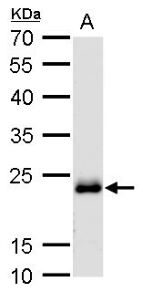
- Western blot analysis of RAB6A using 50 µg mouse brain lysate. Samples were loaded onto a 12% SDS-PAGE gel and probed with a RAB6A polyclonal antibody (Product # PA5-22127) at a dilution of 1:1000.

- Submitted by
- Invitrogen Antibodies (provider)
- Main image

- Experimental details
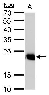
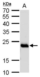
- Western blot analysis of RAB6A using 50 µg rat brain cell lysate. Samples were loaded onto a 12% SDS-PAGE gel and probed with a RAB6A polyclonal antibody (Product # PA5-22127) at a dilution of 1:1000.
- Submitted by
- Invitrogen Antibodies (provider)
- Main image

- Experimental details
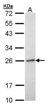
- Western blot analysis of RAB6A using 30 µg of H1299 lysate. Samples were loaded onto a 12% SDS-PAGE gel and probed with a RAB6A polyclonal antibody (Product # PA5-22127) at a dilution of 1:1000.
- Submitted by
- Invitrogen Antibodies (provider)
- Main image
- Experimental details
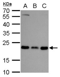
- Western Blot analysis of RAB6A was performed by separating 30 µg of various whole cell lysates by 12 % SDS-PAGE. Proteins were transferred to a membrane and probed with a RAB6A Polyclonal Antibody (Product # PA5-22127) at a dilution of 1:1000. A. A549, B. H1299 , C. HCT116.

- Submitted by
- Invitrogen Antibodies (provider)
- Main image

- Experimental details
- Western Blot analysis of RAB6A was performed by separating 50 µg of mouse brain extracts by 12 % SDS-PAGE. Proteins were transferred to a membrane and probed with a RAB6A Polyclonal Antibody (Product # PA5-22127) at a dilution of 1:1000.
- Submitted by
- Invitrogen Antibodies (provider)
- Main image
- Experimental details
- Western Blot analysis of RAB6A was performed by separating 50 µg of rat brain extracts by 12 % SDS-PAGE. Proteins were transferred to a membrane and probed with a RAB6A Polyclonal Antibody (Product # PA5-22127) at a dilution of 1:1000.

Supportive validation
- Submitted by
- Invitrogen Antibodies (provider)
- Main image
- Experimental details
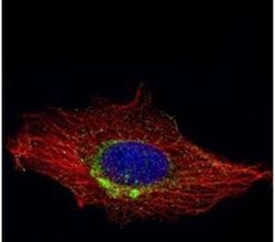
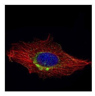
- Immunofluorescent analysis of RAB6A in methanol-fixed HeLa cells using a RAB6A polyclonal antibody (Product # PA5-22127) (Green) at a 1:1000 dilution. Alpha-tubulin filaments were labeled with Product # PA5-29281 (Red) at a 1:2000.

- Submitted by
- Invitrogen Antibodies (provider)
- Main image

- Experimental details
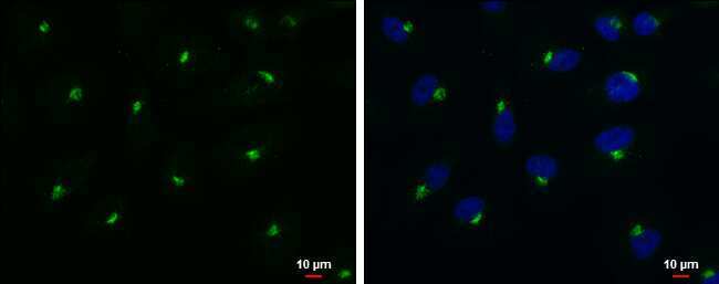
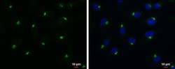
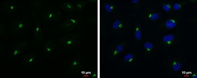
- Immunocytochemistry-Immunofluorescence analysis of RAB6A was performed in A549 cells fixed in 4% paraformaldehyde at RT for 15 min. Green: RAB6A Polyclonal Antibody (Product # PA5-22127) diluted at 1:500. Blue: Hoechst 33342 staining. Scale bar = 10 µm.
- Submitted by
- Invitrogen Antibodies (provider)
- Main image
- Experimental details
- Immunocytochemistry-Immunofluorescence analysis of RAB6A was performed in A549 cells fixed in 4% paraformaldehyde at RT for 15 min. Green: RAB6A Polyclonal Antibody (Product # PA5-22127) diluted at 1:500. Blue: Hoechst 33342 staining. Scale bar = 10 µm.
- Submitted by
- Invitrogen Antibodies (provider)
- Main image

- Experimental details
- Confocal immunofluorescence analysis (Olympus FV10i) of methanol-fixed HeLa, using RAB6A antibody (Product # PA5-22127) (Green) at 1:1,000 dilution. Alpha-tubulin filaments were labeled with (Product # MA1-25054) (Red) at 1:2,000.
Supportive validation
- Submitted by
- Invitrogen Antibodies (provider)
- Main image
- Experimental details
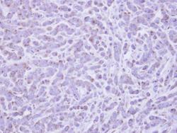
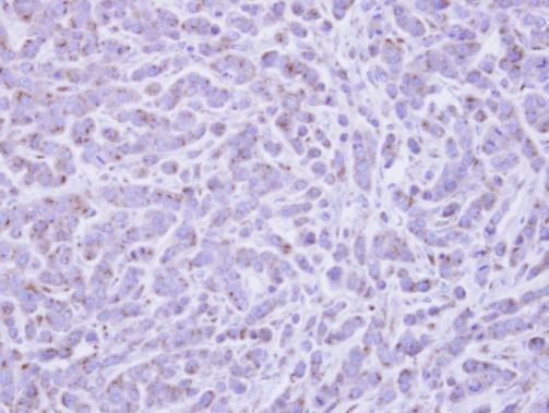
- Immunohistochemical analysis of paraffin-embedded MDA-MB-468 xenograft, using RAB6A (Product # PA5-22127) antibody at 1:500 dilution. Antigen Retrieval: EDTA based buffer, pH 8.0, 15 min.
Supportive validation
- Submitted by
- Invitrogen Antibodies (provider)
- Main image
- Experimental details
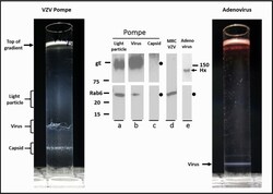
- FIG 6 Purification of virus from Pompe cells by density gradient sedimentation. Monolayers of VZV-infected Pompe cells were harvested by procedures described in Materials and Methods and then subjected to density gradient sedimentation to purify light particles and viral particles (left side). After completion of sedimentation, the gradient was photographed and the individual bands were removed for analysis by immunoblotting. As a control for VZV particles purified by gradient sedimentation, adenovirus was also purified in identical sedimentation gradients and photographed, and the single viral band was removed for analysis by immunoblotting (right side). (Blot a) VZV light particle band immunoblotted for gE and Rab6. Molecular weight markers are included alongside this lane. (Blot b) VZV virus band immunoblotted for gE and Rab6. (Blot c) VZV capsid band immunoblotted for gE and Rab6. (Blot d) VZV-infected cell lysate immunoblotted for Rab6. (Blot e) Adenovirus band immunoblotted for viral hexon (Hx) and Rab6.
