 Explore
 Validate
 Learn
Learn Western blot
Antibody data
- Antibody Data
- Antigen structure
- References [5]
- Comments [0]
- Validations
- Western blot [2]
- Immunocytochemistry [2]
- Immunoprecipitation [1]
- Immunohistochemistry [1]
- Chromatin Immunoprecipitation [1]
Submit
Validation data
Reference
Comment
Report error
- Product number
- GTX102226 - Provider product page

- Provider
- GeneTex
- Proper citation
- GeneTex Cat#GTX102226, RRID:AB_2037324
- Product name
- KAP1 antibody [N3C2], Internal
- Antibody type
- Polyclonal
- Reactivity
- Human, Mouse
- Host
- Rabbit
Submitted references MRI Is a DNA Damage Response Adaptor during Classical Non-homologous End Joining.
BET Protein BRDT Complexes With HDAC1, PRMT5, and TRIM28 and Functions in Transcriptional Repression During Spermatogenesis.
Mass spectrometry-based quantification of the cellular response to methyl methanesulfonate treatment in human cells.
KAP-1 promotes resection of broken DNA ends not protected by γ-H2AX and 53BP1 in G₁-phase lymphocytes.
Functional intersection of ATM and DNA-dependent protein kinase catalytic subunit in coding end joining during V(D)J recombination.
Hung PJ, Johnson B, Chen BR, Byrum AK, Bredemeyer AL, Yewdell WT, Johnson TE, Lee BJ, Deivasigamani S, Hindi I, Amatya P, Gross ML, Paull TT, Pisapia DJ, Chaudhuri J, Petrini JJH, Mosammaparast N, Amarasinghe GK, Zha S, Tyler JK, Sleckman BP
Molecular cell 2018 Jul 19;71(2):332-342.e8
Molecular cell 2018 Jul 19;71(2):332-342.e8
BET Protein BRDT Complexes With HDAC1, PRMT5, and TRIM28 and Functions in Transcriptional Repression During Spermatogenesis.
Wang L, Wolgemuth DJ
Journal of cellular biochemistry 2016 Jun;117(6):1429-38
Journal of cellular biochemistry 2016 Jun;117(6):1429-38
Mass spectrometry-based quantification of the cellular response to methyl methanesulfonate treatment in human cells.
Aslanian A, Yates JR 3rd, Hunter T
DNA repair 2014 Mar;15:29-38
DNA repair 2014 Mar;15:29-38
KAP-1 promotes resection of broken DNA ends not protected by γ-H2AX and 53BP1 in G₁-phase lymphocytes.
Tubbs AT, Dorsett Y, Chan E, Helmink B, Lee BS, Hung P, George R, Bredemeyer AL, Mittal A, Pappu RV, Chowdhury D, Mosammaparast N, Krangel MS, Sleckman BP
Molecular and cellular biology 2014 Aug;34(15):2811-21
Molecular and cellular biology 2014 Aug;34(15):2811-21
Functional intersection of ATM and DNA-dependent protein kinase catalytic subunit in coding end joining during V(D)J recombination.
Lee BS, Gapud EJ, Zhang S, Dorsett Y, Bredemeyer A, George R, Callen E, Daniel JA, Osipovich O, Oltz EM, Bassing CH, Nussenzweig A, Lees-Miller S, Hammel M, Chen BP, Sleckman BP
Molecular and cellular biology 2013 Sep;33(18):3568-79
Molecular and cellular biology 2013 Sep;33(18):3568-79
No comments: Submit comment
Supportive validation
- Submitted by
- GeneTex (provider)
- Main image
- Experimental details
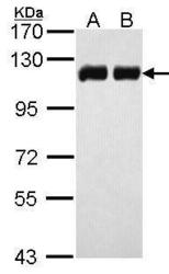
- Sample (30 ?g of whole cell lysate) A: H1299 B: HeLa 7.5% SDS PAGE GTX102226 diluted at 1:10000The HRP-conjugated anti-rabbit IgG antibody (GTX213110-01) was used to detect the primary antibody.

- Submitted by
- GeneTex (provider)
- Main image

- Experimental details
- Sample (30 ?g of whole cell lysate) A:NIH-3T37.5% SDS PAGE GTX102226 diluted at 1:1000 The HRP-conjugated anti-rabbit IgG antibody (GTX213110-01) was used to detect the primary antibody.

Supportive validation
- Submitted by
- GeneTex (provider)
- Main image
- Experimental details
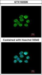
- Immunofluorescence analysis of paraformaldehyde-fixed A431, using KAP1(GTX102226) antibody at 1:200 dilution.

- Submitted by
- GeneTex (provider)
- Main image
- Experimental details
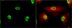
- KAP1 antibody [N3C2], Internal detects KAP1 protein at nucleus by immunofluorescent analysis.Sample: HeLa cells were fixed in 4% paraformaldehyde at RT for 15 min.Green: KAP1 protein stained by KAP1 antibody [N3C2], Internal (GTX102226) diluted at 1:200.Red: alpha Tubulin, a cytoskeleton marker, stained by alpha Tubulin antibody [B-5-1-2] (GTX11304) diluted at 1:10000.

Supportive validation
- Submitted by
- GeneTex (provider)
- Main image

- Experimental details
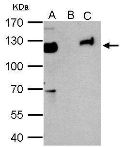
- KAP1 antibody immunoprecipitates KAP1 protein in IP experiments. IP Sample: 1000 ?g HeLa whole cell lysate/extract A. 50 £gg HeLa whole cell lysate/extract B. Control with 2 £gg of preimmune rabbit IgG C. Immunoprecipitation of KAP1 protein by 2 £gg of KAP1 antibody (GTX102226) 7.5% SDS-PAGE The immunoprecipitated KAP1 protein was detected by KAP1 antibody (GTX102226) diluted at 1:1000. EasyBlot anti-rabbit IgG (GTX221666-01) was used as a secondary reagent.
Supportive validation
- Submitted by
- GeneTex (provider)
- Main image
- Experimental details
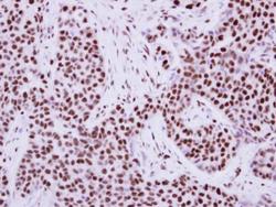
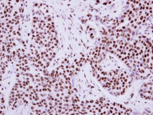
- Immunohistochemical analysis of paraffin-embedded human breast cancer, using KAP1(GTX102226) antibody at 1:500 dilution.
Supportive validation
- Submitted by
- GeneTex (provider)
- Main image

- Experimental details
- ChIP assay followed by QPCR on a known KAP1-binding regionwithin the ZNF180 3¡¦UTR (Mol Cell Biol, 2011, Lyengar et al)
