 Explore
 Validate
 Learn
Learn Western blot
Antibody data
- Antibody Data
- Antigen structure
- References [0]
- Comments [0]
- Validations
- Western blot [2]
- Immunocytochemistry [1]
- Immunohistochemistry [1]
- Flow cytometry [1]
Submit
Validation data
Reference
Comment
Report error
- Product number
- ASR-011-200UL - Provider product page

- Provider
- Invitrogen Antibodies
- Product name
- S1PR1 (EDG1) (extracellular) Polyclonal Antibody
- Antibody type
- Polyclonal
- Antigen
- Other
- Description
- Reconstitution: 1 X 25 µL double distilled water (DDW), depending on the sample size. The antibody ships as a lyophilized powder at room temperature. Upon arrival, it should be stored at -20C. The reconstituted solution can be stored at 4C for up to 1 week. For longer periods, small aliquots should be stored at -20C. Avoid multiple freezing and thawing. Centrifuge all antibody preparations before use (10000 x g 5 min).
- Reactivity
- Human, Mouse, Rat
- Host
- Rabbit
- Isotype
- IgG
- Vial size
- 200 µL
- Concentration
- 0.75 mg/mL
- Storage
- -20° C, Avoid Freeze/Thaw Cycles
No comments: Submit comment
Supportive validation
- Submitted by
- Invitrogen Antibodies (provider)
- Main image

- Experimental details
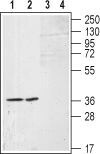
- Western blot analysis of (RAEC) rat aortic endothelial cell line lysates (lanes 1 and 3) and A-10 Rat thoracic aorta smooth muscle (lane 2 and 4) cell lysates: - 1,2. Anti-S1PR1 (EDG1) (extracellular) Antibody (#ASR-011), (1:200).3,4. Anti-S1PR1 (EDG1) (extracellular) Antibody , preincubated with S1PR1/EDG1 (extracellular) Blocking Peptide (#BLP-SR011).

- Submitted by
- Invitrogen Antibodies (provider)
- Main image

- Experimental details
- Western blot analysis of (RAEC) rat aortic endothelial cell line lysates (lanes 1 and 3) and A-10 Rat thoracic aorta smooth muscle (lane 2 and 4) cell lysates: - 1,2. Anti-S1PR1 (EDG1) (extracellular) Antibody (#ASR-011), (1:200).3,4. Anti-S1PR1 (EDG1) (extracellular) Antibody , preincubated with S1PR1/EDG1 (extracellular) Blocking Peptide (#BLP-SR011).
Supportive validation
- Submitted by
- Invitrogen Antibodies (provider)
- Main image

- Experimental details
- Expression of Sphingosine 1-phosphate receptor 1 in mouse 3T3 cells - Cell surface detection of Sphingosine 1-phosphate receptor 1 inmouse live 3T3 cells. A. Extracellular staining of cells using Anti-S1PR1 (EDG1) (extracellular) Antibody (#ASR-011), (1:50), (red). B. DAPI is used for nuclear staining (blue). Merged image of A and B.

Supportive validation
- Submitted by
- Invitrogen Antibodies (provider)
- Main image

- Experimental details
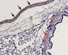
- Expression of Sphingosine 1-phosphate receptor 1 in rat lung - Immunohistochemical staining of paraffin embedded rat lung sections using Anti-S1PR1 (EDG1) (extracellular) Antibody (#ASR-011), (1:100). Staining is present in vascular smooth muscle (black arrows) but not in the muscular layer of bronchi (red arrows). Hematoxilin is used as the counterstain.
Supportive validation
- Submitted by
- Invitrogen Antibodies (provider)
- Main image
- Experimental details
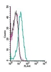
- Cell surface detection of Sphingosine 1-phosphate receptor 1 by indirect flow cytometry in live intact human Jurkat T-cell leukemia cells: - (black line) cells. (red) Cells + goat- Anti-rabbit-APC. (green)Cells + Anti-S1PR1 (EDG1) (extracellular) Antibody (#ASR-011), (5μg) + goat- Anti-rabbit-APC.
