 Explore
 Validate
 Learn
Learn Western blot
Antibody data
- Antibody Data
- Antigen structure
- References [0]
- Comments [0]
- Validations
- Western blot [2]
- Immunohistochemistry [2]
- Flow cytometry [2]
Submit
Validation data
Reference
Comment
Report error
- Product number
- PA5-17938 - Provider product page

- Provider
- Invitrogen Antibodies
- Product name
- SOCS1 Polyclonal Antibody
- Antibody type
- Polyclonal
- Antigen
- Synthetic peptide
- Description
- This antibody is predicted to react with bovine, canine and rat based on sequence homology. This antibody is tested in Peptide ELISA: antibody detection limit dilution 4,000.
- Reactivity
- Human, Mouse
- Host
- Goat
- Isotype
- IgG
- Vial size
- 100 µg
- Concentration
- 0.5 mg/mL
- Storage
- -20° C, Avoid Freeze/Thaw Cycles
No comments: Submit comment
Supportive validation
- Submitted by
- Invitrogen Antibodies (provider)
- Main image

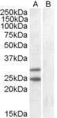
- Experimental details
- Western blot of HEK293 overexpressing SOCS1 using Product # PA5-17938, mock transfection as a control in second lane.
- Submitted by
- Invitrogen Antibodies (provider)
- Main image
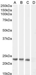
- Experimental details
- Western blot analysis of SOCS1 by a SOCS1 monoclonal antibody (Product # PA5-17938) at a concentration of 0.3 µg/mL. Human Spelen (A), Pig Pancreas (B), (1 µg/mL) Mouse Spleen (C) lysate and negative control HeLa (D) cell lysate (35µg protein in RIPA buffer). Detected by chemiluminescence.

Supportive validation
- Submitted by
- Invitrogen Antibodies (provider)
- Main image
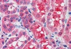
- Experimental details
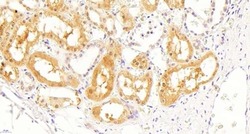
- Immunohistochemical staining of paraffin embedded of Human Kidney using Product # PA5-17938 at a concentration of 2.5 µg/mL. The tissue was processed by steamed antigen retrieval with citrate buffer pH 6 and stained with AP.
- Submitted by
- Invitrogen Antibodies (provider)
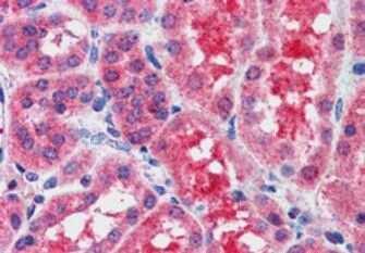
- Main image
- Experimental details
- Immunohistochemical analysis of SOCS1 in Human Kidney using a SOCS1 monoclonal antibody (Product #PA5-17938) at 5 µg/mL. The Human Kidney tissue section was paraffin embeded and detected using Heat induced antigen retrieval with citrate buffer pH 6, HRP-staining.

Supportive validation
- Submitted by
- Invitrogen Antibodies (provider)
- Main image
- Experimental details
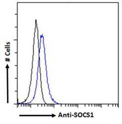
- Flow cytometric analysis of SOCS1 in HEK293 cells using a SOCS1 monoclonal antibody (Product # PA5-17938) at 10 µg/mL for 1hr, depicted by the blue line. The cells were paraformaldehyde fixed and permeabilized with 0.5% Triton. Primary incubation followed by Alexa Fluor 488 secondary antibody (1 µg/mL). IgG control: Unimmunized goat IgG (black line) followed by Alexa Fluor 488 secondary antibody.

- Submitted by
- Invitrogen Antibodies (provider)
- Main image
- Experimental details
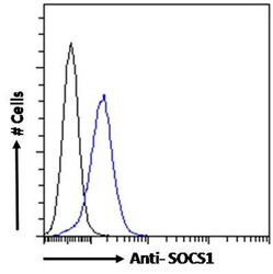
- Flow cytometric analysis of SOCS1 in HEK293 cells using a polyclonal antibody (Product #PA5-17938). HEK293 cells (blue line) were paraformaldehyde fixed and permeabilized with 0.5% Triton. The primary antibody was incubated for one hour (10 µg/mL) followed by an Alexa Fluor 488 secondary antibody (1 µg/mL). IgG control: Unimmunized goat IgG (black line) followed by an Alexa Fluor 488 secondary antibody.
