 Explore
 Validate
 Learn
Learn Western blot
Antibody data
- Antibody Data
- Antigen structure
- References [0]
- Comments [0]
- Validations
- Western blot [2]
- Immunocytochemistry [1]
- Immunohistochemistry [7]
- Flow cytometry [1]
Submit
Validation data
Reference
Comment
Report error
- Product number
- UM500009 - Provider product page

- Provider
- OriGene
- Proper citation
- OriGene Cat#UM500009, RRID:AB_2629024
- Product name
- CD5 mouse monoclonal antibody, clone UMAB9
- Antibody type
- Monoclonal
- Description
- CD5 mouse monoclonal antibody, clone UMAB9
- Host
- Mouse
- Conjugate
- Unconjugated
- Epitope
- CD5
- Isotype
- IgG
- Antibody clone number
- UMAB9
- Vial size
- 100 µl
- Concentration
- 1.00mg/ml
No comments: Submit comment
Supportive validation
- Submitted by
- OriGene (provider)
- Main image

- Experimental details
- Western blot analysis of extracts (35ug) from 9 different cell lines by using anti-CD5 monoclonal antibody (Clone UMAB9) at 1:500.
- Validation comment
- WB

- Submitted by
- OriGene (provider)
- Main image

- Experimental details
- Western Blot analysis of 10 different human tissue lysates (10ug) by using anti-CD5 monoclonal antibody (clone UMAB9, 1:500)
- Validation comment
- WB

Supportive validation
- Submitted by
- OriGene (provider)
- Main image

- Experimental details
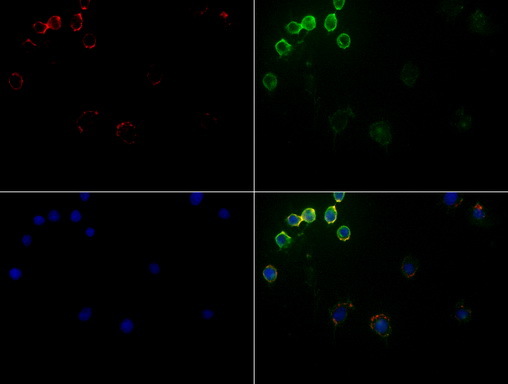
- Immunofluorescent staining of Jurkat cells using CD5 mouse monoclonal antibody (UM500009, green). Actin filaments were labeled with TRITC-phalloidin (red), and nuclear with DAPI (blue). The three-color overlay image is located at the bottom-right corner.
- Validation comment
- IF
Supportive validation
- Submitted by
- OriGene (provider)
- Main image
- Experimental details
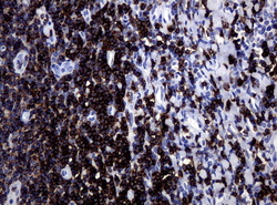
- Immunohistochemical staining of paraffin-embedded Human tonsil using anti-CD5 mouse monoclonal antibody. (Clone UMAB9, dilution 1:100; heat-induced epitope retrieval by 10mM citric buffer, pH6.0, 120C for 3min)

- Submitted by
- OriGene (provider)
- Main image

- Experimental details
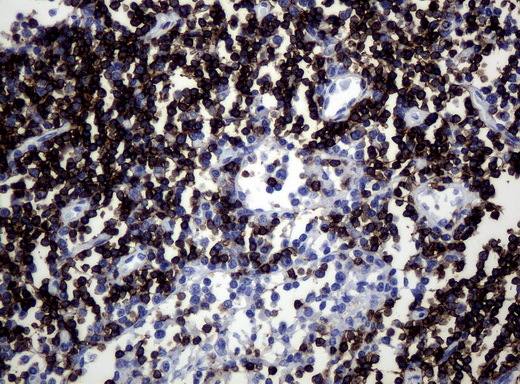
- Immunohistochemical staining of paraffin-embedded Human lymphoma tissue using anti-CD5 mouse monoclonal antibody. (Clone UMAB9, dilution 1:100; heat-induced epitope retrieval by 10mM citric buffer, pH6.0, 120C for 3min)
- Validation comment
- IHC

- Submitted by
- OriGene (provider)
- Main image

- Experimental details
- Immunohistochemical staining of paraffin-embedded lymph node tissue using anti-CD5 mouse monoclonal antibody. (Clone UMAB9, dilution 1:100; heat-induced epitope retrieval by 10mM citric buffer, pH6.0, 120C for 3min)
- Submitted by
- OriGene (provider)
- Main image

- Experimental details
- Immunohistochemical staining of paraffin-embedded human tonsil using anti-CD5 clone UMAB9 mouse monoclonal antibody at 1:200 dilution of 0.1mg/mL detection with Polink2 Broad HRP DAB (UM500009 requires heat-induced epitope retrieval with Citrate pH6.0 at 95-100C 20 minutes. The image shows strong membranous and cytoplasmic staining in >75 % of non germinal center cells of tonsil and
- Validation comment
- IHC

- Submitted by
- OriGene (provider)
- Main image

- Experimental details
- Immunohistochemical staining of paraffin-embedded mouse thymus tissue using anti-CD5 clone UMAB9 mouse monoclonal antibody. (Heat-induced epitope retrieval by 1mM EDTA in 10mM Tris buffer (pH8.5) at 120°C for 3 min, UM500009)(1:300).
- Validation comment
- IHC

- Submitted by
- OriGene (provider)
- Main image

- Experimental details
- Immunohistochemical staining of paraffin-embedded mouse spleen tissue using anti-CD5 clone UMAB9 mouse monoclonal antibody.(Heat-induced epitope retrieval by 1mM EDTA in 10mM Tris buffer (pH8.5) at 120 oC for 3 min, UM500009)(1:300).
- Validation comment
- IHC

- Submitted by
- OriGene (provider)
- Main image

- Experimental details
- Immunohistochemical staining of paraffin-embedded mouse ascending colon tissue using anti-CD5 clone UMAB9 mouse monoclonal antibody. (Heat-induced epitope retrieval by 1mM EDTA in 10mM Tris buffer (pH8.5) at 120°C for 3 min, UM500009)(1:300).
- Validation comment
- IHC

Supportive validation
- Submitted by
- OriGene (provider)
- Main image
- Experimental details
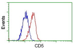
- Flow cytometric Analysis of Jurkat cells, using anti-CD5 antibody(UM500009),(Red), compared to a nonspecific negative control antibody,(Blue).
- Validation comment
- FC
