Explore
 Validate
 Learn
Learn Western blot
 ELISA
ELISAAntibody data
- Antibody Data
- Antigen structure
- References [1]
- Comments [0]
- Validations
- Western blot [1]
- Immunohistochemistry [1]
Submit
Validation data
Reference
Comment
Report error
- Product number
- NB120-7616 - Provider product page

- Provider
- Novus Biologicals
- Proper citation
- Novus Cat#NB120-7616, RRID:AB_788054
- Product name
- Goat Polyclonal Apolipoprotein B/ApoB Antibody
- Antibody type
- Polyclonal
- Description
- Immunogen affinity purified. Typically less than 1% cross reactivity against other types of apoLipoprotein was detected by ELISA. This reacts with human apoLipoprotein A-II and has negligible cross-reactivity with Type A-I, A-II, C-I, C-II, C-III, E and J apoLipoproteins.
- Reactivity
- Human
- Host
- Goat
- Isotype
- IgG
- Vial size
- 0.2 mg
- Concentration
- 1 mg/ml
- Storage
- Store at 4C short term. For extended storage, add an equal volume of glycerol, aliquot and store at -20C or below. Avoid repeated freeze-thaw cycles.
Submitted references Plasma Nitration of High-Density and Low-Density Lipoproteins in Chronic Kidney Disease Patients Receiving Kidney Transplants.
Bakillah A, Tedla F, Ayoub I, John D, Norin AJ, Hussain MM, Brown C
Mediators of inflammation 2015;2015:352356
Mediators of inflammation 2015;2015:352356
No comments: Submit comment
Supportive validation
- Submitted by
- Novus Biologicals (provider)
- Main image
- Experimental details
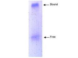
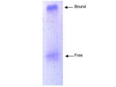
- Western Blot: Apolipoprotein B/ApoB Antibody [NB120-7616] - Coommassie stained gel showing both free and HDL bound Apolipoprotein B/ApoB eluted from a solid phase resin prepared using anti-Human Apolipoprotein B/ApoB antibody. The resin was reacted with human serum prior to washing and elution of bound proteins. The gel was composed of 0.75% agarose in a native buffer system. Separation occurred at room temperature.
Supportive validation
- Submitted by
- Novus Biologicals (provider)
- Main image
- Experimental details
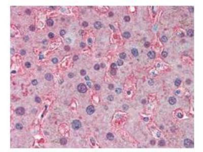
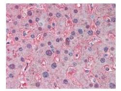
- Immunohistochemistry-Paraffin: Apolipoprotein B/ApoB Antibody [NB120-7616] - Apolipoprotein B/ApoB antibody was used at a 5 ug/ml to detect signal in human liver tissue. Tissue was formalin-fixed and paraffin embedded.