Explore
 Validate
 Learn
LearnANR-051-200UL
antibody from Invitrogen Antibodies
Targeting: NECTIN1
CD111, CLPED1, ED4, HIgR, HVEC, OFC7, PRR, PRR1, PVRL1, PVRR1, SK-12
 Western blot
Antibody data
- Antibody Data
- Antigen structure
- References [0]
- Comments [0]
- Validations
- Western blot [3]
- Immunocytochemistry [1]
- Immunohistochemistry [1]
- Flow cytometry [1]
Submit
Validation data
Reference
Comment
Report error
- Product number
- ANR-051-200UL - Provider product page

- Provider
- Invitrogen Antibodies
- Product name
- Nectin-1/PVRL1 (extracellular) Polyclonal Antibody
- Antibody type
- Polyclonal
- Antigen
- Other
- Description
- Reconstitution: 1 X 25 µL double distilled water (DDW), depending on the sample size. The antibody ships as a lyophilized powder at room temperature. Upon arrival, it should be stored at -20C. The reconstituted solution can be stored at 4C for up to 1 week. For longer periods, small aliquots should be stored at -20C. Avoid multiple freezing and thawing. Centrifuge all antibody preparations before use (10000 x g 5 min).
- Reactivity
- Human, Mouse, Rat
- Host
- Rabbit
- Isotype
- IgG
- Vial size
- 200 µL
- Concentration
- 0.8 mg/mL
- Storage
- -20° C, Avoid Freeze/Thaw Cycles
No comments: Submit comment
Supportive validation
- Submitted by
- Invitrogen Antibodies (provider)
- Main image

- Experimental details
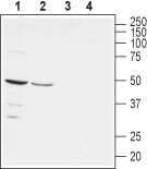
- Western blot analysis of rat (lanes 1 and 3) and mouse (lanes 2 and 4) brain lysates: - 1,2. Anti-Nectin-1/PVRL1 (extracellular) Antibody (#ANR-051), (1:200).3,4. Anti-Nectin-1/PVRL1 (extracellular) Antibody , preincubated with Nectin-1/PVRL1 (extracellular) Blocking Peptide (#BLP-NR051).
- Submitted by
- Invitrogen Antibodies (provider)
- Main image

- Experimental details
- Western blot analysis of rat (lanes 1 and 3) and mouse (lanes 2 and 4) brain lysates: - 1,2. Anti-Nectin-1/PVRL1 (extracellular) Antibody (#ANR-051), (1:200).3,4. Anti-Nectin-1/PVRL1 (extracellular) Antibody , preincubated with Nectin-1/PVRL1 (extracellular) Blocking Peptide (#BLP-NR051).

- Submitted by
- Invitrogen Antibodies (provider)
- Main image
- Experimental details
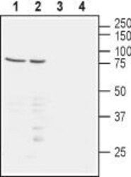
- Western blot analysis of human Jurkat T-cell leukemia (lanes 1 and 3) and human MEG-01 chronic myelogenous leukemia (lanes 2 and 4) cell lysates: - 1,2. Anti-Nectin-1/PVRL1 (extracellular) Antibody (#ANR-051), (1:200).3,4. Anti-Nectin-1/PVRL1 (extracellular) Antibody , preincubated with Nectin-1/PVRL1 (extracellular) Blocking Peptide (#BLP-NR051).

Supportive validation
- Submitted by
- Invitrogen Antibodies (provider)
- Main image

- Experimental details
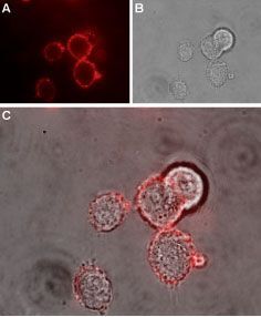
- Expression of Nectin-1 in rat PC12 cells - Cell surface detection of Nectin-1 in live intact rat PC12 pheochromocytoma cells. A. Extracellular staining of cells with Anti-Nectin-1/PVRL1 (extracellular) Antibody (#ANR-051), (1:100), followed by goat Anti-rabbit-AlexaFluor-594secondary Antibody (red). B. Live image of the cells. C. Merge of the two images.
Supportive validation
- Submitted by
- Invitrogen Antibodies (provider)
- Main image

- Experimental details
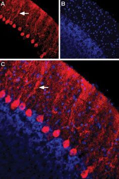
- Expression of Nectin-1 in rat cerebellum - Immunohistochemical staining of immersion-fixed, free floating rat brain frozen sections using Anti-Nectin-1/PVRL1 (extracellular) Antibody (#ANR-051), (1:100). A. Nectin-1staining (red) is apparent in Purkinje neurons and their dendritic tree (arrow). B. Cell nuclei in the same section are visualized with DAPI (blue). C. Merge of the two images.
Supportive validation
- Submitted by
- Invitrogen Antibodies (provider)
- Main image
- Experimental details
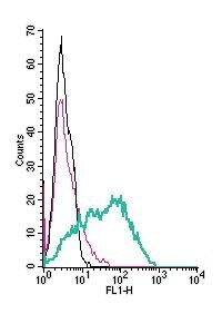
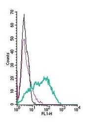
- Cell surface detection of Nectin-1 by indirect flow cytometry in live intact human MEG-01 megakaryoblastic leukemia cells: - (black line) cells. (red) Cells + goat- Anti-rabbit-FITC. (green) Cells + Anti-Nectin-1/PVRL1 (extracellular) Antibody (#ANR-051), (2.5μg) + goat- Anti-rabbit-FITC.