 Explore
 Validate
 Learn
Learn Western blot
Antibody data
- Antibody Data
- Antigen structure
- References [3]
- Comments [0]
- Validations
- Western blot [1]
- Immunocytochemistry [3]
- Immunohistochemistry [1]
Submit
Validation data
Reference
Comment
Report error
- Product number
- AF5005 - Provider product page

- Provider
- R&D Systems
- Product name
- Human/Mouse Cited-2 Antibody
- Antibody type
- Polyclonal
- Description
- Antigen Affinity-purified. Detects mouse Cited-2 in Western blots.
- Reactivity
- Human, Mouse
- Host
- Sheep
- Conjugate
- Unconjugated
- Antigen sequence
O35740- Isotype
- IgG
- Vial size
- 100 ug
- Concentration
- LYOPH
- Storage
- Use a manual defrost freezer and avoid repeated freeze-thaw cycles. 12 months from date of receipt, -20 to -70 °C as supplied. 1 month, 2 to 8 °C under sterile conditions after reconstitution. 6 months, -20 to -70 °C under sterile conditions after reconstitution.
Submitted references Cited2, a transcriptional modulator protein, regulates metabolism in murine embryonic stem cells.
HIF-1α deletion partially rescues defects of hematopoietic stem cell quiescence caused by Cited2 deficiency.
Cited2 gene controls pluripotency and cardiomyocyte differentiation of murine embryonic stem cells through Oct4 gene.
Li Q, Hakimi P, Liu X, Yu WM, Ye F, Fujioka H, Raza S, Shankar E, Tang F, Dunwoodie SL, Danielpour D, Hoppel CL, Ramírez-Bergeron DL, Qu CK, Hanson RW, Yang YC
The Journal of biological chemistry 2014 Jan 3;289(1):251-63
The Journal of biological chemistry 2014 Jan 3;289(1):251-63
HIF-1α deletion partially rescues defects of hematopoietic stem cell quiescence caused by Cited2 deficiency.
Du J, Chen Y, Li Q, Han X, Cheng C, Wang Z, Danielpour D, Dunwoodie SL, Bunting KD, Yang YC
Blood 2012 Mar 22;119(12):2789-98
Blood 2012 Mar 22;119(12):2789-98
Cited2 gene controls pluripotency and cardiomyocyte differentiation of murine embryonic stem cells through Oct4 gene.
Li Q, Ramírez-Bergeron DL, Dunwoodie SL, Yang YC
The Journal of biological chemistry 2012 Aug 17;287(34):29088-100
The Journal of biological chemistry 2012 Aug 17;287(34):29088-100
No comments: Submit comment
Supportive validation
- Submitted by
- R&D Systems (provider)
- Main image

- Experimental details
- Detection of Human/Mouse Cited-2 by Western Blot. Western blot shows lysates of HeLa human cervical epithelial carcinoma cell line, C2C12 mouse myoblast cell line, and NIH-3T3 mouse embryonic fibroblast cell line. PVDF membrane was probed with 1 µg/mL of Sheep Anti-Human/Mouse Cited-2 Antigen Affinity-purified Polyclonal Antibody (Catalog # AF5005) followed by HRP-conjugated Anti-Sheep IgG Secondary Antibody (Catalog # HAF016). A specific band was detected for Cited-2 at approximately 30 kDa (as indicated). This experiment was conducted under reducing conditions and using Immunoblot Buffer Group 1.

Supportive validation
- Submitted by
- R&D Systems (provider)
- Main image

- Experimental details
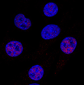
- Cited-2 in HeLa Human Cell Line. Cited-2 was detected in immersion fixed HeLa human cervical epithelial carcinoma cell line using Sheep Anti-Human/Mouse Cited-2 Antigen Affinity-purified Polyclonal Antibody (Catalog # AF5005) at 1 µg/mL for 3 hours at room temperature. Cells were stained using the NorthernLights™ 557-conjugated Anti-Sheep IgG Secondary Antibody (red; Catalog # NL010) and counterstained with DAPI (blue). Specific staining was localized to nuclei. View our protocol for Fluorescent ICC Staining of Cells on Coverslips.

- Submitted by
- R&D Systems (provider)
- Main image

- Experimental details
- Cited-2 in C2C12 Mouse Cell Line. Cited-2 was detected in immersion fixed C2C12 mouse myoblast cell line using Sheep Anti-Human/Mouse Cited-2 Antigen Affinity-purified Polyclonal Antibody (Catalog # AF5005) at 1 µg/mL for 3 hours at room temperature. Cells were stained using the NorthernLights™ 557-conjugated Anti-Sheep IgG Secondary Antibody (red; Catalog # NL010) and counterstained with DAPI (blue). Specific staining was localized to nuclei. View our protocol for Fluorescent ICC Staining of Cells on Coverslips.
- Submitted by
- R&D Systems (provider)
- Main image

- Experimental details
- Cited-2 in NIH-3T3 Mouse Cell Line. Cited-2 was detected in immersion fixed NIH-3T3 mouse embryonic fibroblast cell line using Sheep Anti-Human/Mouse Cited-2 Antigen Affinity-purified Polyclonal Antibody (Catalog # AF5005) at 1 µg/mL for 3 hours at room temperature. Cells were stained using the NorthernLights™ 557-conjugated Anti-Sheep IgG Secondary Antibody (red; Catalog # NL010) and counterstained with DAPI (blue). Specific staining was localized to nuclei. View our protocol for Fluorescent ICC Staining of Cells on Coverslips.

Supportive validation
- Submitted by
- R&D Systems (provider)
- Main image

- Experimental details
- Cited-2 in Human Breast Cancer Tissue. Cited-2 was detected in immersion fixed paraffin-embedded sections of human breast cancer tissue using Sheep Anti-Human/Mouse Cited-2 Antigen Affinity-purified Polyclonal Antibody (Catalog # AF5005) at 3 µg/mL overnight at 4 °C. Tissue was stained using the Anti-Sheep HRP-DAB Cell & Tissue Staining Kit (brown; Catalog # CTS019) and counterstained with hematoxylin (blue). Specific staining was localized to nuclei. View our protocol for Chromogenic IHC Staining of Paraffin-embedded Tissue Sections.
