Explore
 Validate
 Learn
Learn Western blot
Antibody data
- Antibody Data
- Antigen structure
- References [1]
- Comments [0]
- Validations
- Western blot [3]
- Immunocytochemistry [2]
- Immunohistochemistry [3]
- Flow cytometry [1]
- Chromatin Immunoprecipitation [1]
- Other assay [1]
Submit
Validation data
Reference
Comment
Report error
- Product number
- MA1-114 - Provider product page

- Provider
- Invitrogen Antibodies
- Product name
- Phospho-CREB/ATF1 (Ser133, Ser63) Monoclonal Antibody (10E9)
- Antibody type
- Monoclonal
- Antigen
- Synthetic peptide
- Description
- This antibody recognizes CREB protein phosphorylated at serine 133 and ATF1 phosphorylated at serine 63. The antibody was purified from serum-free cell culture supernatant by subsequent thiophilic adsorption and size exclusion chromatography.
- Antibody clone number
- 1.00E+10
- Concentration
- 0.1 mg/mL
Submitted references protaTETHER - a method for the incorporation of variable linkers in protein fusions reveals impacts of linker flexibility in a PKAc-GFP fusion protein.
Norris JL, Hughes RM
FEBS open bio 2018 Jun;8(6):1029-1042
FEBS open bio 2018 Jun;8(6):1029-1042
No comments: Submit comment
Supportive validation
- Submitted by
- Invitrogen Antibodies (provider)
- Main image

- Experimental details
- Western blot analysis of Phospho-CREB pSer133 was performed by loading 20 µg of untreated HeLa cells (lane 1) and HeLa cells treated with 200 nM PMA (lane 2) onto an SDS polyacrylamide gel. Proteins were transferred to a PVDF membrane and blocked at 4ºC overnight. The membrane was probed with a Phospho-CREB pSer133 monoclonal antibody (Product # MA1-114) at a dilution of 1:500 overnight at 4°C, washed in TBST, and probed with an HRP-conjugated secondary antibody for 1 hr at room temperature in the dark. Chemiluminescent detection was performed using Pierce ECL Plus Western Blotting Substrate (Product # 32132). Results show a band at ~43 kDa.

- Submitted by
- Invitrogen Antibodies (provider)
- Main image

- Experimental details
- Western blot analysis of Phospho CREB performed by using nuclear extract of PMA treated HELA cells. Cell were starved for 6 -8 hours with DMEM containing 1% FBS and treated with 200 nM of PMA for 5 & 20 minutes. Nuclear extracts were prepared using NE-PER kit (Product # 78833). Western blot analysis was done by loading ~ 30 ugs of nuclear extract and 15 µL of PageRuler Prestained Protein Ladder (Product # 26616) per well onto a 4-20% Tris-HCl polyacrylamide gel. Proteins were transferred to a PVDF membrane using the G2 Fast Blotter (Product # 62288) and blocked with StartingBlock (PBS) Blocking Buffer (Product # 37538) for at least 1 hour at room temperature. Phospho CREB was detected at ~43 kD using a Phoso CREB monoclonal antibody (Product # MA1-114) at a concentration of 4 µg/mL in blocking buffer overnight at 4C on a rocking platform, followed by an HRP-conjugated goat anti-mouse IgG (light chain specific) secondary antibody at a dilution of 1:10,000 for at least 1 hour. Chemiluminescent detection was performed using Super Signal West Dura (Product # 34075).

- Submitted by
- Invitrogen Antibodies (provider)
- Main image
- Experimental details
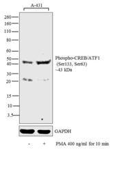
- Western blot analysis was performed on whole cell extracts (30 µg lysate) of A-431 (Lane 1) and A-431 treated with PMA (400 ng/mL for 10 min) (Lane 2). The blot was probed with Anti-Phospho-Phospho-CREB/ATF1 (Ser133, Ser63) Monoclonal Antibody (Product # MA1-114, 1:500 dilution) and detected by chemiluminescence using Goat anti-Mouse IgG (H+L) Superclonal™ Secondary Antibody, HRP conjugate (Product # A28177, 0.25 µg/mL, 1:4000 dilution). A 43 kDa band corresponding to Phospho-CREB/ATF1 (Ser133, Ser63) was detected in untreated lysate and increased upon PMA treatment.

Supportive validation
- Submitted by
- Invitrogen Antibodies (provider)
- Main image
- Experimental details
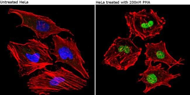
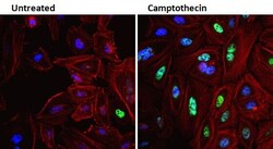
- Immunofluorescent analysis of Phospho-CREB pSer133 (green) showing staining in the in the nucleus of untreated HeLa cells (left) and HeLa cells treated with PMA (right) (right) compared to a negative control without primary antibody (left). Formalin-fixed cells were permeabilized with 0.1% Triton X-100 in TBS for 5-10 minutes and blocked with 3% BSA-PBS for 30 minutes at room temperature. Cells were probed with a Phospho-CREB pSer133 monoclonal antibody (Product # MA1-114) in 3% BSA-PBS at a dilution of 1:100 and incubated overnight at 4ºC in a humidified chamber. Cells were washed with PBST and incubated with a DyLight-conjugated secondary antibody in PBS at room temperature in the dark. F-actin (red) was stained with a fluorescent red phalloidin and nuclei (blue) were stained with Hoechst or DAPI. Images were taken at a magnification of 60x.
- Submitted by
- Invitrogen Antibodies (provider)
- Main image
- Experimental details
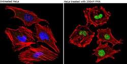
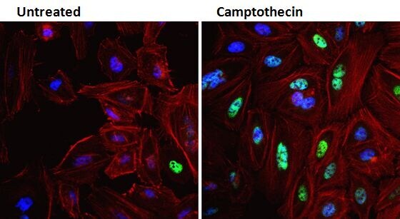
- Immunofluorescent analysis of Phospho-CREB pSer133 (green) in HeLa cells either left untreated (left panel) or treated with 1uM Camptothecin (right panel) for 20 hours. Formalin fixed cells were permeabilized with 0.1% Triton X-100 in TBS for 10 minutes at room temperature and blocked with 1% Blocker BSA (Product # 37525) for 15 minutes at room temperature. Cells were probed with a Phospho-CREB pSer133 monoclonal antibody (Product # MA1-114) at a concentration of 4 µg/mL for at least 1 hour at room temperature, washed with PBS, and incubated with DyLight 488 goat anti-mouse IgG secondary antibody (Product # 35502) at a dilution of 1:400 for 30 minutes at room temperature. F-Actin (red) was stained with DyLight 554 Phalloidin (Product # 21834) and nuclei (blue) were stained with Hoechst 33342 dye (Product # 62249). Images were taken on a Thermo Scientific ArrayScan and ToxInsight Instrument at 20X magnification.
Supportive validation
- Submitted by
- Invitrogen Antibodies (provider)
- Main image
- Experimental details
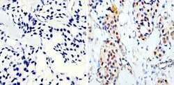
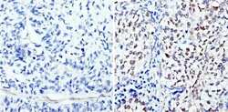
- Immunohistochemistry analysis of Phospho-CREB pSer133 showing staining in the nucleus of paraffin-embedded human breast carcinoma (right) compared to a negative control without primary antibody (left). To expose target proteins, antigen retrieval was performed using 10mM sodium citrate (pH 6.0), microwaved for 8-15 min. Following antigen retrieval, tissues were blocked in 3% H2O2-methanol for 15 min at room temperature, washed with ddH2O and PBS, and then probed with a Phospho-CREB pSer133 monoclonal antibody (Product # MA1-114) diluted in 3% BSA-PBS at a dilution of 1:200 overnight at 4°C in a humidified chamber. Tissues were washed extensively in PBST and detection was performed using an HRP-conjugated secondary antibody followed by colorimetric detection using a DAB kit. Tissues were counterstained with hematoxylin and dehydrated with ethanol and xylene to prep for mounting.
- Submitted by
- Invitrogen Antibodies (provider)
- Main image
- Experimental details
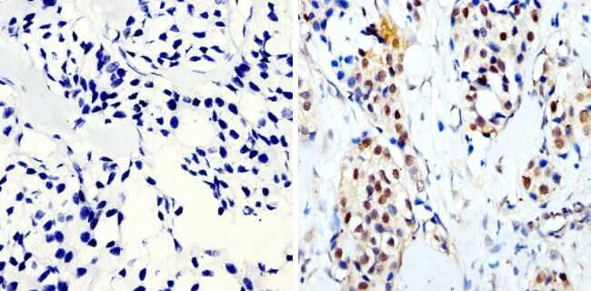
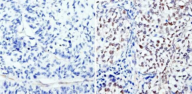
- Immunohistochemistry analysis of Phospho-CREB pSer133 showing staining in the nucleus of paraffin-embedded human lung squamous carcinoma (right) compared to a negative control without primary antibody (left). To expose target proteins, antigen retrieval was performed using 10mM sodium citrate (pH 6.0), microwaved for 8-15 min. Following antigen retrieval, tissues were blocked in 3% H2O2-methanol for 15 min at room temperature, washed with ddH2O and PBS, and then probed with a Phospho-CREB pSer133 monoclonal antibody (Product # MA1-114) diluted in 3% BSA-PBS at a dilution of 1:200 overnight at 4°C in a humidified chamber. Tissues were washed extensively in PBST and detection was performed using an HRP-conjugated secondary antibody followed by colorimetric detection using a DAB kit. Tissues were counterstained with hematoxylin and dehydrated with ethanol and xylene to prep for mounting.
- Submitted by
- Invitrogen Antibodies (provider)
- Main image
- Experimental details
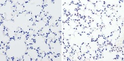
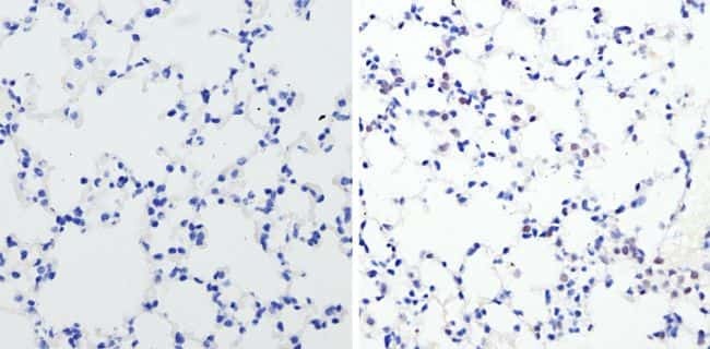
- Immunohistochemistry analysis of Phospho-CREB pSer133 showing staining in the nucleus of paraffin-embedded mouse lung tissue (right) compared to a negative control without primary antibody (left). To expose target proteins, antigen retrieval was performed using 10mM sodium citrate (pH 6.0), microwaved for 8-15 min. Following antigen retrieval, tissues were blocked in 3% H2O2-methanol for 15 min at room temperature, washed with ddH2O and PBS, and then probed with a Phospho-CREB pSer133 monoclonal antibody (Product # MA1-114) diluted in 3% BSA-PBS at a dilution of 1:100 overnight at 4°C in a humidified chamber. Tissues were washed extensively in PBST and detection was performed using an HRP-conjugated secondary antibody followed by colorimetric detection using a DAB kit. Tissues were counterstained with hematoxylin and dehydrated with ethanol and xylene to prep for mounting.
Supportive validation
- Submitted by
- Invitrogen Antibodies (provider)
- Main image
- Experimental details
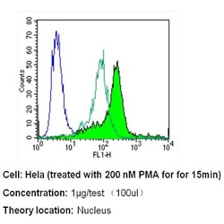
- Flow cytometry analysis of Phospho-CREB pSer133 in untreated Hela cells (open green histogram) and Hela cells treated with 200 nM PMA for for 15 min (filled green histogram) compared to an isotype control (blue). Cells were harvested, adjusted to a concentration of 1-5x10^6 cells/mL, fixed with 2% paraformaldehyde and washed with PBS. Cells were blocked with a 2% solution of BSA-PBS for 30 min at room temperature and incubated with a Phospho-CREB pSer133 monoclonal antibody (Product # MA1-114) at a dilution of 1 µg/test for 40 min at room temperature. Cells were then incubated for 40 min at room temperature in the dark using a Dylight 488-conjugated secondary antibody and re-suspended in PBS for FACS analysis.

Supportive validation
- Submitted by
- Invitrogen Antibodies (provider)
- Main image
- Experimental details
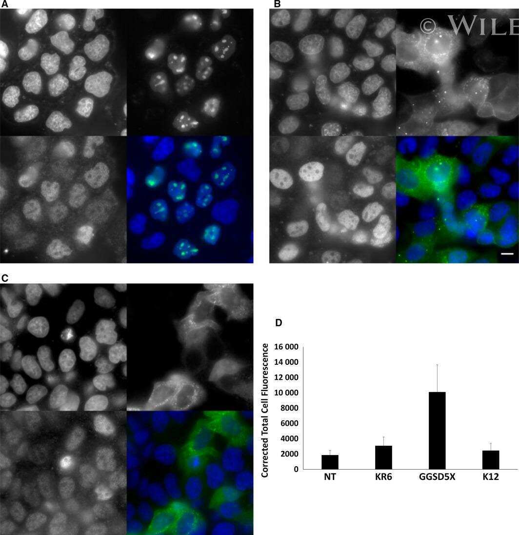
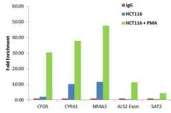
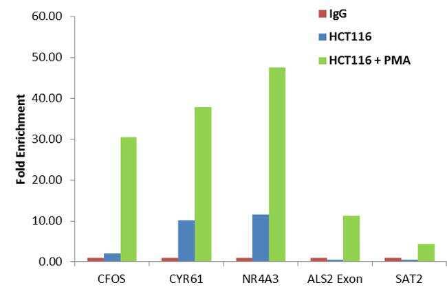
- Enrichment of endogenous Phospho-CREB/ATF1 (Ser133, Ser63) protein at specific gene loci using Anti-Phospho-CREB/ATF1 (Ser133, Ser63) Antibody: Chromatin Immunoprecipitation (ChIP) was performed using Anti-Phospho-CREB/ATF1 (Ser133, Ser63) Mouse Monoclonal Antibody (Product # MA1-114, 10 ul) on sheared chromatin from 2 million control HCT 116 and serum-starved PMA-treated HCT 116 cells using the MAGnify ChIP system kit (Product # 49-2024). Normal Rabbit IgG was used as a negative IP control. The purified DNA was analyzed by qPCR with PCR primer pairs over the cFOS, CYR61, NR4A3, ALS2 genes (active) and SAT2 satellite repeats (inactive). Data is presented as fold enrichment of the antibody signal in control and treated cells versus the negative control IgG using the comparative CT method.
Supportive validation
- Submitted by
- Invitrogen Antibodies (provider)
- Main image
- Experimental details
- NULL