Explore
 Validate
 Learn
Learn Western blot
Antibody data
- Antibody Data
- Antigen structure
- References [1]
- Comments [0]
- Validations
- Western blot [2]
- Immunocytochemistry [1]
- Immunohistochemistry [1]
- Flow cytometry [1]
- Other assay [1]
Submit
Validation data
Reference
Comment
Report error
- Product number
- MA5-15597 - Provider product page

- Provider
- Invitrogen Antibodies
- Product name
- GSK3B Monoclonal Antibody (3D10)
- Antibody type
- Monoclonal
- Antigen
- Purifed from natural sources
- Description
- MA5-15597 targets GSK3B in FACS, IF, IHC, and WB applications and shows reactivity with Human, mouse, Non-human primate, and Rat samples.
- Antibody clone number
- 3D10
- Concentration
- Conc. Not Determined
Submitted references Proteomic characterization of post-mortem human brain tissue following ultracentrifugation-based subcellular fractionation.
Kandigian SE, Ethier EC, Kitchen RR, Lam TT, Arnold SE, Carlyle BC
Brain communications 2022;4(3):fcac103
Brain communications 2022;4(3):fcac103
No comments: Submit comment
Supportive validation
- Submitted by
- Invitrogen Antibodies (provider)
- Main image

- Experimental details
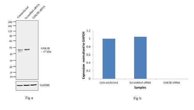
- Knockdown of GSK3B was achieved by transfecting MCF7 cells with GSK3B specific validated siRNAs (Silencer® select Product # S6240). Western blot analysis (Fig. a) was performed using whole cell extracts from the GSK3B knockdown cells (lane 3), non-specific scrambled siRNA transfected cells (lane 2) and untransfected cells (lane 1). The blots were probed with GSK3B Monoclonal Antibody (Product # MA5-15597, 1:500 dilution) and Goat anti-Mouse IgG (H+L) Superclonal™ Secondary Antibody, HRP conjugate (Product # A28177, 0.25 µg/mL, 1:4000 dilution). Densitometric analysis of this western blot is shown in histogram (Fig b). Decrease in signal upon siRNA mediated knock down confirms that antibody is specific to GSK3B.
- Submitted by
- Invitrogen Antibodies (provider)
- Main image

- Experimental details
- Western blot analysis was performed on whole cell extracts of MCF7 (Lane 1), MDA-MB-231 (Lane 2), HeLa (Lane 3), Neuro-2a (Lane 4), U-87 MG (Lane 5), A-431 ( Lane 6) and NIH/3T3 (Lane 7). The blot was probed with Anti-GSK3B antibody (Product # MA5-15597, 1:500 dilution) and detected by chemiluminescence using Goat anti-Mouse IgG (H+L) Superclonal™ Secondary Antibody, HRP conjugate (Product # A28177, 0.25 µg/mL, 1:4000 dilution). A 47 kDa band corresponding to 4EBP1 was observed across the cell lines tested.

Supportive validation
- Submitted by
- Invitrogen Antibodies (provider)
- Main image
- Experimental details
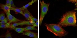
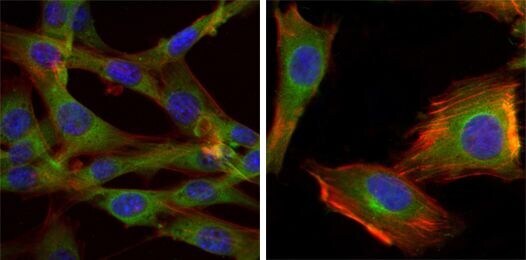
- Immunofluorescence analysis of NIH/3T3 (left) and U251 (right) cells using GSK3B monoclonal antibody (Product # MA5-15597) (Green). Blue: DRAQ5 fluorescent DNA dye. Red: actin filaments have been labeled with phalloidin.
Supportive validation
- Submitted by
- Invitrogen Antibodies (provider)
- Main image

- Experimental details
- Immunohistochemical analysis of paraffin-embedded human lung cancer (left) and breast cancer tissues (right) using GSK3B monoclonal antibody (Product # MA5-15597) followed with DAB staining.

Supportive validation
- Submitted by
- Invitrogen Antibodies (provider)
- Main image
- Experimental details
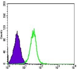
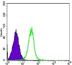
- Flow cytometric analysis of HeLa cells using GSK3B monoclonal antibody (Product # MA5-15597) (green) and negative control (purple).
Supportive validation
- Submitted by
- Invitrogen Antibodies (provider)
- Main image
- Experimental details
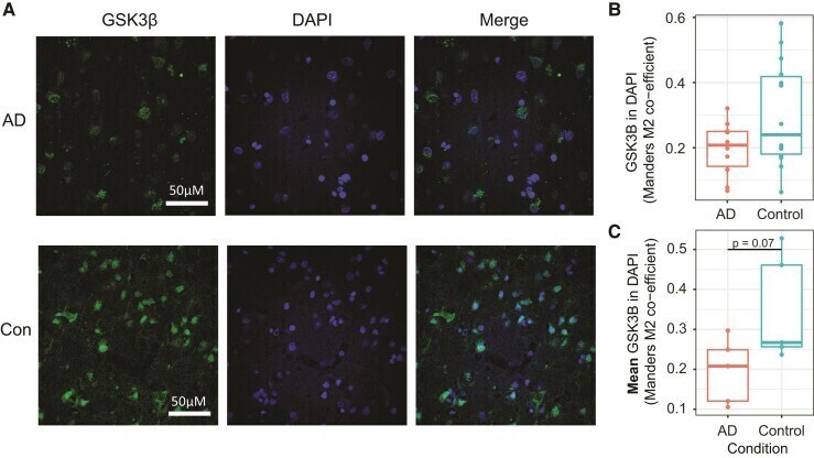
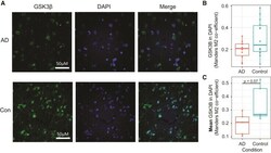
- Immunohistochemistry of angular gyrus sections with anti-GSK3beta shows a trend towards increased nuclear GSK3beta in controls . ( A ) Representative images show increased colocalization of GSK3beta signal with nuclear DAPI staining. Subjective visual analysis suggests increased presence of nuclear speckles in Control samples compared with AD. ( B ) Plot showing Mander's M2 overlap coefficient for each individual image shows an enrichment for increased nuclear overlap of GSK3beta staining with DAPI. ( C ) The mean Mander's M2 coefficient for each subject shows a trend (Student's t -test, T = -2.18, P = 0.07) towards increased nuclear overlap of GSK3beta in controls compared with AD.