Explore
 Validate
 Learn
Learn Western blot
 Immunoprecipitation
Antibody data
- Antibody Data
- Antigen structure
- References [3]
- Comments [0]
- Validations
- Western blot [1]
Submit
Validation data
Reference
Comment
Report error
- Product number
- AF5430 - Provider product page

- Provider
- R&D Systems
- Product name
- Mouse Complement Factor D/Adipsin Antibody
- Antibody type
- Polyclonal
- Description
- Immunogen affinity purified. Detects mouse Complement Factor D/Adipsin in direct ELISAs and Western blots. In direct ELISAs, approximately 10% cross-reactivity with recombinant human (rh) Complement Factor D is observed and less than 5% cross-reactivity with rhGranzyme A is observed.
- Reactivity
- Mouse
- Host
- Sheep
- Conjugate
- Unconjugated
- Antigen sequence
P03953- Isotype
- IgG
- Vial size
- 100 ug
- Concentration
- LYOPH
- Storage
- Use a manual defrost freezer and avoid repeated freeze-thaw cycles. 12 months from date of receipt, -20 to -70 °C as supplied. 1 month, 2 to 8 °C under sterile conditions after reconstitution. 6 months, -20 to -70 °C under sterile conditions after reconstitution.
Submitted references Contribution of Adipose-Derived Factor D/Adipsin to Complement Alternative Pathway Activation: Lessons from Lipodystrophy.
Regulation of C3 Activation by the Alternative Complement Pathway in the Mouse Retina.
Complement factor B is the downstream effector of TLRs and plays an important role in a mouse model of severe sepsis.
Wu X, Hutson I, Akk AM, Mascharak S, Pham CTN, Hourcade DE, Brown R, Atkinson JP, Harris CA
Journal of immunology (Baltimore, Md. : 1950) 2018 Apr 15;200(8):2786-2797
Journal of immunology (Baltimore, Md. : 1950) 2018 Apr 15;200(8):2786-2797
Regulation of C3 Activation by the Alternative Complement Pathway in the Mouse Retina.
Williams JA, Stampoulis D, Gunter CE, Greenwood J, Adamson P, Moss SE
PloS one 2016;11(8):e0161898
PloS one 2016;11(8):e0161898
Complement factor B is the downstream effector of TLRs and plays an important role in a mouse model of severe sepsis.
Zou L, Feng Y, Li Y, Zhang M, Chen C, Cai J, Gong Y, Wang L, Thurman JM, Wu X, Atkinson JP, Chao W
Journal of immunology (Baltimore, Md. : 1950) 2013 Dec 1;191(11):5625-35
Journal of immunology (Baltimore, Md. : 1950) 2013 Dec 1;191(11):5625-35
No comments: Submit comment
Supportive validation
- Submitted by
- R&D Systems (provider)
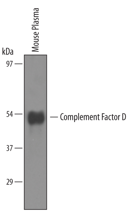
- Main image
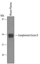
- Experimental details
- Detection of Mouse Complement Factor D/Adipsin by Western Blot. Western blot shows lysates of mouse plasma. PVDF membrane was probed with 1 µg/mL of Sheep Anti-Mouse Complement Factor D/Adipsin Antigen Affinity-purified Polyclonal Antibody (Catalog # AF5430) followed by HRP-conjugated Anti-Sheep IgG Secondary Antibody (Catalog # HAF016). A specific band was detected for Complement Factor D/Adipsin at approximately 45kDa (as indicated). This experiment was conducted under reducing conditions and using Immunoblot Buffer Group 8.