Explore
 Validate
 Learn
Learn Western blot
Antibody data
- Antibody Data
- Antigen structure
- References [0]
- Comments [0]
- Validations
- Western blot [3]
- Immunohistochemistry [2]
Submit
Validation data
Reference
Comment
Report error
- Product number
- PA5-22307 - Provider product page

- Provider
- Invitrogen Antibodies
- Product name
- CD41 Polyclonal Antibody
- Antibody type
- Polyclonal
- Antigen
- Recombinant protein fragment
- Description
- Recommended positive controls: A549, mouse spleen. Predicted reactivity: Mouse (89%), Dog (88%), Pig (82%), Rabbit (87%), Bovine (86%). PA5-22307 detects ITGA2b (CD41) protein in formalin-fixed paraffin-embedded equine endometrial tissue. Store product as a concentrated solution. Centrifuge briefly prior to opening the vial.
- Reactivity
- Human, Mouse
- Host
- Rabbit
- Isotype
- IgG
- Vial size
- 100 µL
- Concentration
- 0.25 mg/mL
- Storage
- Store at 4°C short term. For long term storage, store at -20°C, avoiding freeze/thaw cycles.
No comments: Submit comment
Supportive validation
- Submitted by
- Invitrogen Antibodies (provider)
- Main image

- Experimental details
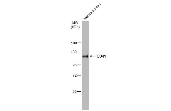
- Western Blot analysis of CD41 was performed by separating 50 µg of Mouse tissue extracts by 7.5% SDS-PAGE. Proteins were transferred to a membrane and probed with a CD41 Polyclonal Antibody (Product # PA5-22307) at a dilution of 1:500. The HRP-conjugated anti-rabbit IgG antibody was used to detect the primary antibody.
- Submitted by
- Invitrogen Antibodies (provider)
- Main image
- Experimental details
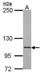
- Western Blot using CD41 Polyclonal Antibody (Product # PA5-22307). Sample (30 µg of whole cell lysate). Lane A: A549. 7.5% SDS PAGE. CD41 Polyclonal Antibody (Product # PA5-22307) diluted at 1:500. The HRP-conjugated anti-rabbit IgG antibody was used to detect the primary antibody.

- Submitted by
- Invitrogen Antibodies (provider)
- Main image

- Experimental details
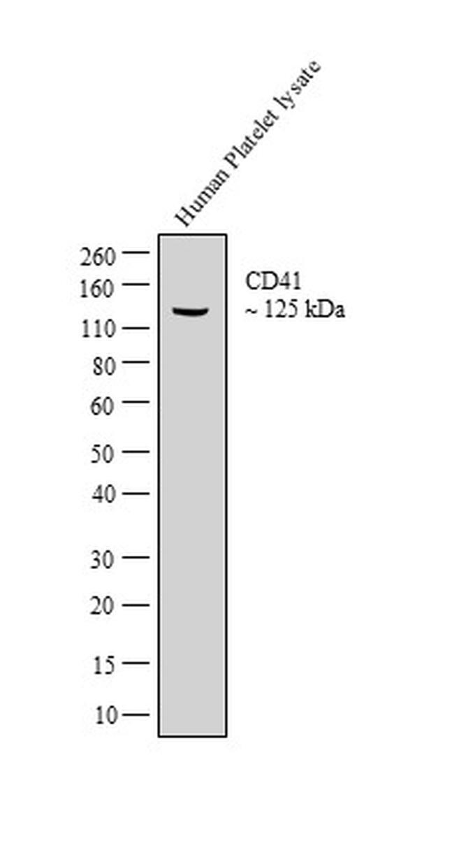
- Western blot analysis was performed on whole cell extracts (30 µg lysate) of Human Platelets (Lane 1). The blot was probed with CD41 Polyclonal Antibody (Product # PA5-22307, 1 µg/mL) and detected by chemiluminescence using Goat anti-Rabbit IgG (H+L) Superclonal™ Secondary Antibody, HRP conjugate (Product # A27036, 0.25 µg/mL, 1:4000 dilution). A 125 kDa band corresponding to CD41 was detected in the lysate tested.
Supportive validation
- Submitted by
- Invitrogen Antibodies (provider)
- Main image
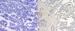
- Experimental details
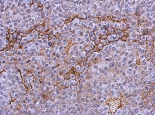
- Immunohistochemistry analysis of CD41 was performed on equine endometrial tissue sections. 5-µm-thick sections were prepared from formalin-fixed paraffin-embedded tissue blocks and were dried on slide plate at 37°C overnight. The sections were placed in a Bond Max Automated Immunohistochemistry Vision Biosystem (Leica Microsystems GmbH, Wetzlar, Germany) according to the following protocol. First, tissues were deparaffinized and pre-treated with Epitope Retrieval Solution 1 (Citrate based pH6) at 98°C for 10 minutes. After washing steps, peroxidase blocking was carried out for 10 min using the Bond Polymer Refine Detection Kit DC9800 (Leica Microsystems GmbH). Tissues were again washed and then incubated with the primary antibody (Product # PA5-22307, 1:200) for 30 min then the secondary antibody diluted 1:1000. Subsequently, tissues were incubated with polymer for 10 min and developed with DAB-Chromogen for 10 min. Data courtesy of Antibody Data Exchange Program.

- Submitted by
- Invitrogen Antibodies (provider)
- Main image

- Experimental details
- Immunohistochemical analysis of paraffin-embedded PC13 xenograft, using CD41 (Product # PA5-22307) antibody at 1:500 dilution. Antigen Retrieval: EDTA based buffer, pH 8.0, 15 min.