Explore
 Validate
 Learn
Learn Western blot
 ELISA
ELISAAntibody data
- Antibody Data
- Antigen structure
- References [3]
- Comments [0]
- Validations
- Western blot [1]
- Immunohistochemistry [1]
Submit
Validation data
Reference
Comment
Report error
- Product number
- NB100-65252 - Provider product page

- Provider
- Novus Biologicals
- Proper citation
- Novus Cat#NB100-65252, RRID:AB_2138456
- Product name
- Rabbit Polyclonal Enolase 1 Antibody
- Antibody type
- Polyclonal
- Description
- Protein G purified. This recognises human Non-neuronal enolase, and demostrates less than 0.1% cross-reactivity with non-specific enolase. Non-neuronal enolase is a glycolytic enzyme composed of homodimeric alpha enolase subunits and is expressed in most human tissues. The gene coding for the 48kDa form of non-neuronal enolase also codes for a shorter 37kDa isoform, myc-binding protein 1 (MBP-1) lacking the N-terminal 93 amino acids of non-neuronal enolase.
- Reactivity
- Human, Bovine
- Host
- Rabbit
- Isotype
- IgG
- Vial size
- 0.5 ml
- Concentration
- 5 mg/ml
- Storage
- Store at 4C short term. Aliquot and store at -20C long term. Avoid freeze-thaw cycles.
Submitted references Hypoxia-Inducible Factor 1α (HIF-1α) Counteracts the Acute Death of Cells Transplanted into the Injured Spinal Cord.
RNA binding protein HuR regulates extracellular matrix gene expression and pH homeostasis independent of controlling HIF-1α signaling in nucleus pulposus cells.
Interaction of arrestin with enolase1 in photoreceptors.
David BT, Curtin JJ, Goldberg DC, Scorpio K, Kandaswamy V, Hill CE
eNeuro 2020 May Jun;7(3)
eNeuro 2020 May Jun;7(3)
RNA binding protein HuR regulates extracellular matrix gene expression and pH homeostasis independent of controlling HIF-1α signaling in nucleus pulposus cells.
Pan H, Strickland A, Madhu V, Johnson ZI, Chand SN, Brody JR, Fertala A, Zheng Z, Shapiro IM, Risbud MV
Matrix biology : journal of the International Society for Matrix Biology 2019 Apr;77:23-40
Matrix biology : journal of the International Society for Matrix Biology 2019 Apr;77:23-40
Interaction of arrestin with enolase1 in photoreceptors.
Smith WC, Bolch S, Dugger DR, Li J, Esquenazi I, Arendt A, Benzenhafer D, McDowell JH
Investigative ophthalmology & visual science 2011 Mar;52(3):1832-40
Investigative ophthalmology & visual science 2011 Mar;52(3):1832-40
No comments: Submit comment
Supportive validation
- Submitted by
- Novus Biologicals (provider)
- Main image

- Experimental details
- Simple Western: Enolase 1 Antibody [NB100-65252] - Simple Western lane view shows a specific band for Enolase 1 in 0.2 mg/ml of HeLa lysate. This experiment was performed under reducing conditions using the 12-230 kDa separation system.

Supportive validation
- Submitted by
- Novus Biologicals (provider)
- Main image

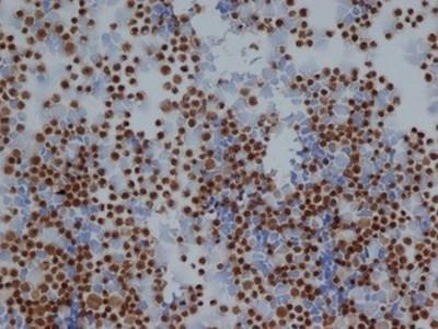
- Experimental details
- Immunohistochemistry: Enolase 1 Antibody [NB100-65252] - Staining of Jurkat cells with rabbit anti human Enolase 1 .