 Explore
 Validate
 Learn
Learn Western blot
Antibody data
- Antibody Data
- Antigen structure
- References [1]
- Comments [0]
- Validations
- Western blot [3]
- Immunocytochemistry [4]
- Immunohistochemistry [2]
- Other assay [1]
Submit
Validation data
Reference
Comment
Report error
- Product number
- MA5-32613 - Provider product page

- Provider
- Invitrogen Antibodies
- Product name
- NEFM Recombinant Rabbit Monoclonal Antibody (JM11-20)
- Antibody type
- Monoclonal
- Antigen
- Synthetic peptide
- Description
- Recombinant rabbit monoclonal antibodies are produced using in vitro expression systems. The expression systems are developed by cloning in the specific antibody DNA sequences from immunoreactive rabbits. Then, individual clones are screened to select the best candidates for production. The advantages of using recombinant rabbit monoclonal antibodies include: better specificity and sensitivity, lot-to-lot consistency, animal origin-free formulations, and broader immunoreactivity to diverse targets due to larger rabbit immune repertoire.
- Reactivity
- Human, Mouse, Rat
- Host
- Rabbit
- Isotype
- IgG
- Antibody clone number
- JM11-20
- Vial size
- 100 µL
- Concentration
- 1 mg/mL
- Storage
- Store at 4°C short term. For long term storage, store at -20°C, avoiding freeze/thaw cycles.
Submitted references Motoneuronal inflammasome activation triggers excessive neuroinflammation and impedes regeneration after sciatic nerve injury.
Molnár K, Nógrádi B, Kristóf R, Mészáros Á, Pajer K, Siklós L, Nógrádi A, Wilhelm I, Krizbai IA
Journal of neuroinflammation 2022 Mar 19;19(1):68
Journal of neuroinflammation 2022 Mar 19;19(1):68
No comments: Submit comment
Supportive validation
- Submitted by
- Invitrogen Antibodies (provider)
- Main image

- Experimental details
- Western blot analysis of NEFM in 293T cells lysates using a NEFM Monoclonal antibody (Product # MA5-32613) at a dilution of 1:500.

- Submitted by
- Invitrogen Antibodies (provider)
- Main image

- Experimental details
- Western blot analysis of NEFM in 293T cells lysates using a NEFM Monoclonal antibody (Product # MA5-32613) at a dilution of 1:500.

- Submitted by
- Invitrogen Antibodies (provider)
- Main image

- Experimental details
- Western blot was performed using Anti-NEFM Recombinant Rabbit Monoclonal Antibody (JM11-20) (Product # MA5-32613) and a 150 kDa band corresponding to NEFM was observed across HEK-293 and MCF7. Whole Cell Extract-WCL (30 µg lysate) of HEK-293 (Lane 1), MCF7 (Lane 2), HeLa (Lane 3), Hep G2 (Lane 4) and K-562 (Lane 5) were electrophoresed using NuPAGE™ 4-12% Bis-Tris Protein Gel (Product # NP0321BOX). Resolved proteins were then transferred onto a Nitrocellulose membrane (Product # IB23001) by iBlot® 2 Dry Blotting System (Product # IB21001). The blot was probed with the primary antibody (1:1000 dilution) and detected by chemiluminescence with Goat anti-Rabbit IgG (H+L) Superclonal™ Recombinant Secondary Antibody, HRP (Product # A27036, 1:4000 dilution) using the iBright FL 1000 (Product # A32752). Chemiluminescent detection was performed using Novex® ECL Chemiluminescent Substrate Reagent Kit (Product # WP20005).

Supportive validation
- Submitted by
- Invitrogen Antibodies (provider)
- Main image

- Experimental details
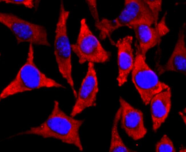
- Immunocytochemical analysis of NEFM in N2A cells using a NEFM Monoclonal antibody (Product # MA5-32613) as seen in red. The nuclear counter stain is DAPI (blue). Cells were fixed in paraformaldehyde, permeabilised with 0.25% Triton X100/PBS.

- Submitted by
- Invitrogen Antibodies (provider)
- Main image

- Experimental details
- Immunocytochemical analysis of NEFM in PC-12 cells using a NEFM Monoclonal antibody (Product # MA5-32613) as seen in red. The nuclear counter stain is DAPI (blue). Cells were fixed in paraformaldehyde, permeabilised with 0.25% Triton X100/PBS.

- Submitted by
- Invitrogen Antibodies (provider)
- Main image

- Experimental details
- Immunocytochemical analysis of NEFM in SH-SY5Y cells using a NEFM Monoclonal antibody (Product # MA5-32613) as seen in red. The nuclear counter stain is DAPI (blue). Cells were fixed in paraformaldehyde, permeabilised with 0.25% Triton X100/PBS.
- Submitted by
- Invitrogen Antibodies (provider)
- Main image

- Experimental details
- Knockout of NEFM was achieved by CRISPR-Cas9 genome editing. Immunofluorescence analysis was performed on wild type HEK-293 cells (panel a,d), HEK-293 Cas9 cells (panels b,e) and HEK-293 NEFM KO cells (panel c,f). Cells were fixed, permeabilized, and labelled with NEFM Recombinant Rabbit Monoclonal Antibody (JM11-20) (Product # MA5-32613) (1:500 dilution), followed by Goat anti-Rabbit IgG (H+L) Highly Cross-Adsorbed Secondary Antibody, Alexa Fluor Plus 488 (Product # A32731) (1:2,000). Nuclei (blue) were stained using ProLong™ Diamond Antifade Mountant with DAPI (Product # P36962), and Rhodamine Phalloidin (Product # R415) (1:300) was used for cytoskeletal F-actin (red) staining. Loss of signal (panel c,f) upon CRISPR mediated knockout (KO) confirms that antibody is specific to NEFM (green). The images were captured at 60X magnification.

Supportive validation
- Submitted by
- Invitrogen Antibodies (provider)
- Main image

- Experimental details
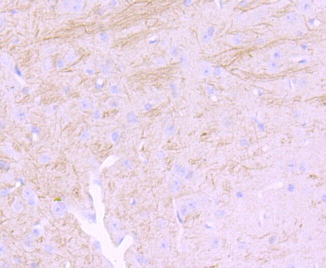
- Immunohistochemical analysis of NEFM of paraffin-embedded Mouse brain tissue using a NEFM Monoclonal antibody (Product #MA5-32613). Counter stained with hematoxylin.
- Submitted by
- Invitrogen Antibodies (provider)
- Main image

- Experimental details
- Immunohistochemical analysis of NEFM of paraffin-embedded rat brain tissue using a NEFM Monoclonal antibody (Product #MA5-32613). Counter stained with hematoxylin.

Supportive validation
- Submitted by
- Invitrogen Antibodies (provider)
- Main image

- Experimental details
- Effect of MCC950 on axonal regrowth 5 days after sciatic nerve axotomy and coaptation. a - d Confocal images of injured sciatic nerves stained with the Schwann cell marker p75 and the axonal marker NEFM. Animals were treated with vehicle ( a ), MCC950 ( b ), goat IgG c or IL-1beta neutralizing antibody ( d ). e Quantification of regrowing axons 5 days after sciatic nerve injury. Bars represent the average number of regrowing axons and their distance relative to the zone of coaptation. N = 3-4 animals/group. f Graph representing the sum of regrowing axons derived as multiplication of axonal length and number of axons (i.e., area under the curve of graph ( e )). Mean values are shown on bars. Bars represent average +- SEM. N = 3-4 animals/group. * p < 0.05 compared to vehicle-treated animals, # p < 0.05 compared to mice receiving goat IgG (Student's paired t-test). Coapt. axotomy + coaptation, IL-1beta neutr. IL-1beta neutralization, a.u. arbitrary unit
