 Explore
 Validate
 Learn
Learn Western blot
Antibody data
- Antibody Data
- Antigen structure
- References [0]
- Comments [0]
- Validations
- Western blot [2]
- Immunohistochemistry [8]
Submit
Validation data
Reference
Comment
Report error
- Product number
- ABIN2506524 - Provider product page

- Provider
- antibodies-online
- Product name
- anti-Testis Expressed 10 (TEX10) (Middle Region) antibody
- Antibody type
- Polyclonal
- Antigen
- The immunogen for Anti-TEX10 antibody is: synthetic peptide directed towards the middle region of Human TEX10
- Description
- Affinity Purified
- Reactivity
- Human, Mouse, Rat, Bovine, Canine, Guinea Pig, Horse, Porcine, Rabbit
- Host
- Rabbit
- Antigen sequence
RLTSQQWRLK VLVRLSKFLQ ALADGSSRLR ES
EGLQEQKE NPHATSNSIF- Epitope
- Middle Region
- Vial size
- 50 μg
- Storage
- Add 50 μL of distilled water. Final anti-TEX10 antibody concentration is 1 mg/mL in PBS buffer with 2 % sucrose. For longer periods of storage, store at -20°C. Avoid repeat freeze-thaw cycles.
- Handling
- Avoid repeat freeze-thaw cycles.
No comments: Submit comment
Supportive validation
- Submitted by
- antibodies-online (provider)
- Main image
- Experimental details
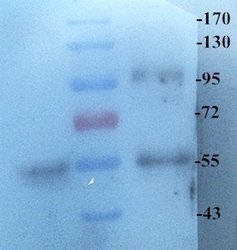
- Western blot analysis of rat lung (left lane), large intestine (right lane) using Cryopyrin antibody (Dilution at 1:500)

- Submitted by
- antibodies-online (provider)
- Main image

- Experimental details
- Western blot analysis of rat lung lysates and human colon carcinoma lysates using Cryopyrin antibody (primary antibody dilution at 1:200)

Supportive validation
- Submitted by
- antibodies-online (provider)
- Main image

- Experimental details
- Immunohistochemical staining of rat skin tissue using Cryopyrin antibody (dilution of primary antibody - 1:200)

- Submitted by
- antibodies-online (provider)
- Main image
- Experimental details
- Immunohistochemical staining of rat skin tissue using Cryopyrin antibody (dilution of primary antibody - 1:200)

- Submitted by
- antibodies-online (provider)
- Main image

- Experimental details
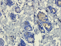
- Immunohistochemical analysis of formalin-fixed and paraffin embedded human colon cancer using Cryopyrin antibody (dilution at 1:200)

- Submitted by
- antibodies-online (provider)
- Main image

- Experimental details
- IHC-P image of pig large intestines tissue using anti-Cryopyrin (dilution of primary antibody at 1:200)
- Submitted by
- antibodies-online (provider)
- Main image

- Experimental details
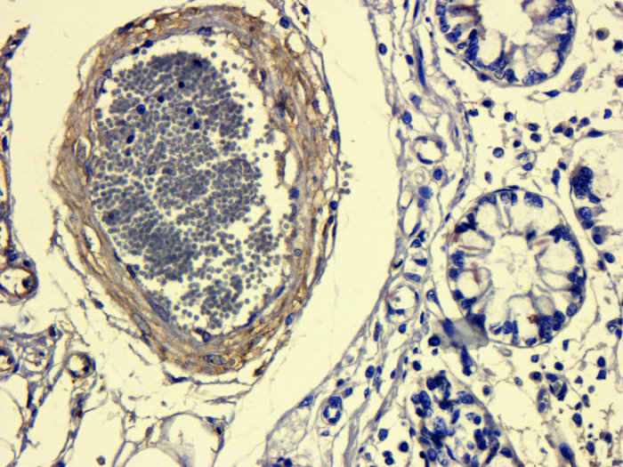
- Immunohistochemical staining of paraffin embedded human thyroid carcinoma tissue using Cryopyrin antibody (primary antibody at 1:200)

- Submitted by
- antibodies-online (provider)
- Main image
- Experimental details
- IHC-P staining of human thyroid carcinoma tissue using anti-Cryopyrin (dilution at 1:200)

- Submitted by
- antibodies-online (provider)
- Main image

- Experimental details
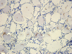
- IHC-P image of rat liver tissue using Cryopyrin antibody (dilution of primary antibody at 1:200)

- Submitted by
- antibodies-online (provider)
- Main image

- Experimental details
- Immunohistochemical staining of rat heart tissue using anti Cryopyrin (dilution of primary antibody - 1:200)
