Explore
 Validate
 Learn
Learn Western blot
Antibody data
- Antibody Data
- Antigen structure
- References [2]
- Comments [0]
- Validations
- Western blot [6]
- Immunocytochemistry [1]
- Immunohistochemistry [1]
Submit
Validation data
Reference
Comment
Report error
- Product number
- GTX100253 - Provider product page

- Provider
- GeneTex
- Proper citation
- GeneTex Cat#GTX100253, RRID:AB_1240648
- Product name
- Cortactin antibody [N1], N-term
- Antibody type
- Polyclonal
- Reactivity
- Human, Mouse, Rat
- Host
- Rabbit
Submitted references Merkel Cell Polyomavirus Small T Antigen Drives Cell Motility via Rho-GTPase-Induced Filopodium Formation.
A study of the spatial protein organization of the postsynaptic density isolated from porcine cerebral cortex and cerebellum.
Stakaitytė G, Nwogu N, Dobson SJ, Knight LM, Wasson CW, Salguero FJ, Blackbourn DJ, Blair GE, Mankouri J, Macdonald A, Whitehouse A
Journal of virology 2018 Jan 15;92(2)
Journal of virology 2018 Jan 15;92(2)
A study of the spatial protein organization of the postsynaptic density isolated from porcine cerebral cortex and cerebellum.
Yun-Hong Y, Chih-Fan C, Chia-Wei C, Yen-Chung C
Molecular & cellular proteomics : MCP 2011 Oct;10(10):M110.007138
Molecular & cellular proteomics : MCP 2011 Oct;10(10):M110.007138
No comments: Submit comment
Supportive validation
- Submitted by
- GeneTex (provider)
- Main image
- Experimental details
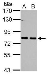
- Sample (30 ug of whole cell lysate) A: NIH-3T3 B: JC 7.5% SDS PAGE GTX100253 diluted at 1:1000

- Submitted by
- GeneTex (provider)
- Main image

- Experimental details
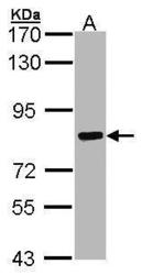
- Sample (30 ug of whole cell lysate) A: PC-12 7.5% SDS PAGE GTX100253 diluted at 1:3000

- Submitted by
- GeneTex (provider)
- Main image
- Experimental details
- Sample (30 ug of whole cell lysate) A: Hep G2 (GTX27900) 7.5% SDS PAGE GTX100253 diluted at 1:1000

- Submitted by
- GeneTex (provider)
- Main image
- Experimental details
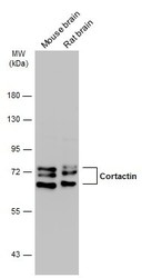
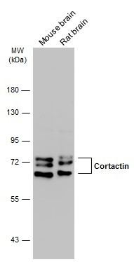
- Various tissue extracts (50 ?g) were separated by 7.5% SDS-PAGE, and the membrane was blotted with Cortactin antibody [N1], N-term (GTX100253) diluted at 1:1000. The HRP-conjugated anti-rabbit IgG antibody (GTX213110-01) was used to detect the primary antibody.
- Submitted by
- GeneTex (provider)
- Main image
- Experimental details
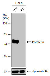
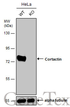
- Wild-type (WT) and Cortactin knockout (KO) HeLa cell extracts (30 ?g) were separated by 7.5% SDS-PAGE, and the membrane was blotted with Cortactin antibody [N1], N-term (GTX100253) diluted at 1:1000. The HRP-conjugated anti-rabbit IgG antibody (GTX213110-01) was used to detect the primary antibody.
- Submitted by
- GeneTex (provider)
- Main image
- Experimental details
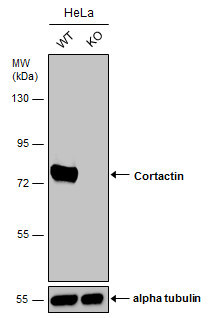
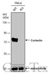
- Wild-type (WT) and Cortactin knockout (KO) HeLa cell extracts (30 ?g) were separated by 7.5% SDS-PAGE, and the membrane was blotted with Cortactin antibody [N1], N-term (GTX100253) diluted at 1:1000. The HRP-conjugated anti-rabbit IgG antibody (GTX213110-01) was used to detect the primary antibody.
Supportive validation
- Submitted by
- GeneTex (provider)
- Main image
- Experimental details
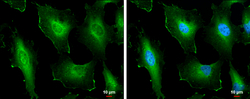
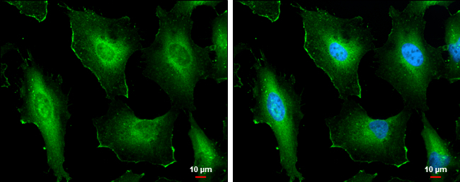
- Cortactin antibody [N1], N-term detects Cortactin protein at cell membrane and cytoplasm by immunofluorescent analysis.Sample: HeLa cells were fixed in 4% paraformaldehyde at RT for 15 min.Green: Cortactin protein stained by Cortactin antibody [N1], N-term (GTX100253) diluted at 1:500.Blue: Hoechst 33342 staining.Scale bar = 10 £gm.
Supportive validation
- Submitted by
- GeneTex (provider)
- Main image
- Experimental details
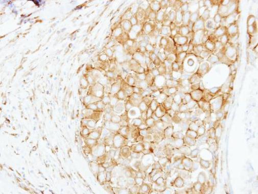
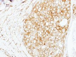
- Immunohistochemical analysis of paraffin-embedded human breast cancer, using Cortactin(GTX100253) antibody at 1:250 dilution.