 Explore
 Validate
 Learn
LearnMA5-14140
antibody from Invitrogen Antibodies
Targeting: L1CAM
CD171, HSAS, HSAS1, MASA, MIC5, S10, SPG1
 Western blot
 Immunoprecipitation
Antibody data
- Antibody Data
- Antigen structure
- References [20]
- Comments [0]
- Validations
- Western blot [4]
- Immunocytochemistry [1]
- Immunohistochemistry [2]
- Other assay [1]
Submit
Validation data
Reference
Comment
Report error
- Product number
- MA5-14140 - Provider product page

- Provider
- Invitrogen Antibodies
- Product name
- CD171 Monoclonal Antibody (UJ127)
- Antibody type
- Monoclonal
- Antigen
- Other
- Description
- MA5-14140 targets L1 Cell Adhesion Molecule in immunohistochemistry (paraffin), immunoprecipitation, and Western blot applications and shows reactivity with human samples. The MA5-14140 immunogen is homogenous suspension of 16 week human fetal brain. MA5-14140 has been successfully used in immunohistochemistry analysis of NCAM1 in mouse E16.5 frozen lung sections.
- Reactivity
- Human, Mouse
- Host
- Mouse
- Isotype
- IgG
- Antibody clone number
- UJ127
- Vial size
- 500 µL
- Concentration
- 0.2 mg/mL
- Storage
- 4° C
Submitted references Targeting FER Kinase Inhibits Melanoma Growth and Metastasis.
Inhibition of Pannexin 1 Reduces the Tumorigenic Properties of Human Melanoma Cells.
Targeting L1 cell adhesion molecule expression using liposome-encapsulated siRNA suppresses prostate cancer bone metastasis and growth.
Inflammatory responses in the muscle coat of stomach and small bowel in the postoperative ileus model of guinea pig.
The dimensions and characteristics of the subepidermal nerve plexus in human skin--terminal Schwann cells constitute a substantial cell population within the superficial dermis.
Adhesion molecule L1 is down-regulated in malignant peripheral nerve sheath tumors versus benign neurofibromatosis type 1-associated tumors.
Two alcohol binding residues interact across a domain interface of the L1 neural cell adhesion molecule and regulate cell adhesion.
Modification of the L1-CAM carboxy-terminus in pancreatic adenocarcinoma cells.
Inside-out regulation of L1 conformation, integrin binding, proteolysis, and concomitant cell migration.
DOG1 and CD117 are the antibodies of choice in the diagnosis of gastrointestinal stromal tumours.
SSEA-1 is an enrichment marker for tumor-initiating cells in human glioblastoma.
Subset of esophageal adenocarcinoma expresses adhesion molecule l1 in contrast to squamous cell carcinoma.
L1 is associated with micrometastatic spread and poor outcome in colorectal cancer.
L1 is associated with favorable outcome in neuroblastomas in contrast to adult tumors.
L1 (CD171) is highly expressed in gastrointestinal stromal tumors.
Identification of cell surface and secreted proteins essential for tumor cell survival using a genetic suppressor element screen.
Extracellular signal-regulated kinase (ERK)-dependent gene expression contributes to L1 cell adhesion molecule-dependent motility and invasion.
Characterization of gene expression in mucinous cystic neoplasms of the pancreas using oligonucleotide microarrays.
Patterning axonal guidance molecules using a novel strategy for microcontact printing.
Patterning axonal guidance molecules using a novel strategy for microcontact printing.
Ivanova IA, Arulanantham S, Barr K, Cepeda M, Parkins KM, Hamilton AM, Johnston D, Penuela S, Hess DA, Ronald JA, Dagnino L
Cancers 2019 Mar 24;11(3)
Cancers 2019 Mar 24;11(3)
Inhibition of Pannexin 1 Reduces the Tumorigenic Properties of Human Melanoma Cells.
Freeman TJ, Sayedyahossein S, Johnston D, Sanchez-Pupo RE, O'Donnell B, Huang K, Lakhani Z, Nouri-Nejad D, Barr KJ, Harland L, Latosinsky S, Grant A, Dagnino L, Penuela S
Cancers 2019 Jan 16;11(1)
Cancers 2019 Jan 16;11(1)
Targeting L1 cell adhesion molecule expression using liposome-encapsulated siRNA suppresses prostate cancer bone metastasis and growth.
Sung SY, Wu IH, Chuang PH, Petros JA, Wu HC, Zeng HJ, Huang WC, Chung LW, Hsieh CL
Oncotarget 2014 Oct 30;5(20):9911-29
Oncotarget 2014 Oct 30;5(20):9911-29
Inflammatory responses in the muscle coat of stomach and small bowel in the postoperative ileus model of guinea pig.
Choi HK, Lee YJ, Lee YH, Park JP, Min K, Park H
Yonsei medical journal 2013 Nov;54(6):1336-41
Yonsei medical journal 2013 Nov;54(6):1336-41
The dimensions and characteristics of the subepidermal nerve plexus in human skin--terminal Schwann cells constitute a substantial cell population within the superficial dermis.
Reinisch CM, Tschachler E
Journal of dermatological science 2012 Mar;65(3):162-9
Journal of dermatological science 2012 Mar;65(3):162-9
Adhesion molecule L1 is down-regulated in malignant peripheral nerve sheath tumors versus benign neurofibromatosis type 1-associated tumors.
Blessmann M, Gröbe A, Quaas A, Kaifi JT, Mistakidis G, Bernreuther C, Sauter G, Gros S, Rawnaq T, Friedrich R, Mautner VF, Smeets R, Heiland M, Schachner M, Izbicki JR
Oral surgery, oral medicine, oral pathology and oral radiology 2012 Feb;113(2):239-44
Oral surgery, oral medicine, oral pathology and oral radiology 2012 Feb;113(2):239-44
Two alcohol binding residues interact across a domain interface of the L1 neural cell adhesion molecule and regulate cell adhesion.
Dou X, Menkari CE, Shanmugasundararaj S, Miller KW, Charness ME
The Journal of biological chemistry 2011 May 6;286(18):16131-9
The Journal of biological chemistry 2011 May 6;286(18):16131-9
Modification of the L1-CAM carboxy-terminus in pancreatic adenocarcinoma cells.
Chen MM, Lee CY, Leland HA, Silletti S
Tumour biology : the journal of the International Society for Oncodevelopmental Biology and Medicine 2011 Apr;32(2):347-57
Tumour biology : the journal of the International Society for Oncodevelopmental Biology and Medicine 2011 Apr;32(2):347-57
Inside-out regulation of L1 conformation, integrin binding, proteolysis, and concomitant cell migration.
Chen MM, Lee CY, Leland HA, Lin GY, Montgomery AM, Silletti S
Molecular biology of the cell 2010 May 15;21(10):1671-85
Molecular biology of the cell 2010 May 15;21(10):1671-85
DOG1 and CD117 are the antibodies of choice in the diagnosis of gastrointestinal stromal tumours.
Novelli M, Rossi S, Rodriguez-Justo M, Taniere P, Seddon B, Toffolatti L, Sartor C, Hogendoorn PC, Sciot R, Van Glabbeke M, Verweij J, Blay JY, Hohenberger P, Flanagan A, Dei Tos AP
Histopathology 2010 Aug;57(2):259-70
Histopathology 2010 Aug;57(2):259-70
SSEA-1 is an enrichment marker for tumor-initiating cells in human glioblastoma.
Son MJ, Woolard K, Nam DH, Lee J, Fine HA
Cell stem cell 2009 May 8;4(5):440-52
Cell stem cell 2009 May 8;4(5):440-52
Subset of esophageal adenocarcinoma expresses adhesion molecule l1 in contrast to squamous cell carcinoma.
Rawnaq T, Kleinhans H, Uto M, Schurr PG, Reichelt U, Cataldegirmen G, Gawad KA, Yekebas EF, Schachner M, Izbicki JR, Kaifi JT
Anticancer research 2009 Apr;29(4):1195-9
Anticancer research 2009 Apr;29(4):1195-9
L1 is associated with micrometastatic spread and poor outcome in colorectal cancer.
Kaifi JT, Reichelt U, Quaas A, Schurr PG, Wachowiak R, Yekebas EF, Strate T, Schneider C, Pantel K, Schachner M, Sauter G, Izbicki JR
Modern pathology : an official journal of the United States and Canadian Academy of Pathology, Inc 2007 Nov;20(11):1183-90
Modern pathology : an official journal of the United States and Canadian Academy of Pathology, Inc 2007 Nov;20(11):1183-90
L1 is associated with favorable outcome in neuroblastomas in contrast to adult tumors.
Wachowiak R, Fiegel HC, Kaifi JT, Quaas A, Krickhahn A, Schurr PG, Erttmann R, Schachner M, Kluth D, Sauter G, Izbicki JR
Annals of surgical oncology 2007 Dec;14(12):3575-80
Annals of surgical oncology 2007 Dec;14(12):3575-80
L1 (CD171) is highly expressed in gastrointestinal stromal tumors.
Kaifi JT, Strelow A, Schurr PG, Reichelt U, Yekebas EF, Wachowiak R, Quaas A, Strate T, Schaefer H, Sauter G, Schachner M, Izbicki JR
Modern pathology : an official journal of the United States and Canadian Academy of Pathology, Inc 2006 Mar;19(3):399-406
Modern pathology : an official journal of the United States and Canadian Academy of Pathology, Inc 2006 Mar;19(3):399-406
Identification of cell surface and secreted proteins essential for tumor cell survival using a genetic suppressor element screen.
Gelman MS, Ye XK, Stull R, Suhy D, Jin L, Ng D, Than B, Ji M, Pan A, Perez P, Sun Y, Yeung P, Garcia LM, Harte R, Lu Y, Lamar E, Tavassoli R, Kennedy S, Osborn S, Chin DJ, Meshaw K, Holzmayer TA, Axenovich SA, Abo A
Oncogene 2004 Oct 21;23(49):8158-70
Oncogene 2004 Oct 21;23(49):8158-70
Extracellular signal-regulated kinase (ERK)-dependent gene expression contributes to L1 cell adhesion molecule-dependent motility and invasion.
Silletti S, Yebra M, Perez B, Cirulli V, McMahon M, Montgomery AM
The Journal of biological chemistry 2004 Jul 9;279(28):28880-8
The Journal of biological chemistry 2004 Jul 9;279(28):28880-8
Characterization of gene expression in mucinous cystic neoplasms of the pancreas using oligonucleotide microarrays.
Fukushima N, Sato N, Prasad N, Leach SD, Hruban RH, Goggins M
Oncogene 2004 Dec 2;23(56):9042-51
Oncogene 2004 Dec 2;23(56):9042-51
Patterning axonal guidance molecules using a novel strategy for microcontact printing.
Oliva AA Jr, James CD, Kingman CE, Craighead HG, Banker GA
Neurochemical research 2003 Nov;28(11):1639-48
Neurochemical research 2003 Nov;28(11):1639-48
Patterning axonal guidance molecules using a novel strategy for microcontact printing.
Oliva AA Jr, James CD, Kingman CE, Craighead HG, Banker GA
Neurochemical research 2003 Nov;28(11):1639-48
Neurochemical research 2003 Nov;28(11):1639-48
No comments: Submit comment
Supportive validation
- Submitted by
- Invitrogen Antibodies (provider)
- Main image
- Experimental details
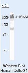
- Western blot of L1 Cell Adhesion Molecule using L1 Cell Adhesion Molecule Monoclonal Antibody (Product # MA5-14140) on IMR-5 Cells.

- Submitted by
- Invitrogen Antibodies (provider)
- Main image
- Experimental details
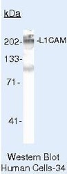
- Western blot of L1 Cell Adhesion Molecule using L1 Cell Adhesion Molecule Monoclonal Antibody (Product # MA5-14140) on IMR-5 Cells.

- Submitted by
- Invitrogen Antibodies (provider)
- Main image
- Experimental details
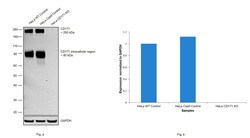
- Knockout of CD171 was achieved by CRISPR-Cas9 genome editing. Western blot analysis of CD171 was performed by loading 20 µg of HeLa wild type (Lane 1), HeLa Cas9 control (Lane 2), HeLa CD171 knockout (Lane 3) membrane enriched cell extracts. The blot was probed with Anti-CD171Monoclonal Antibody (UJ127) (Product # MA5-14140) using 1 µg/mL and Goat anti-Mouse IgG (H+L), Superclonal™ Recombinant Secondary Antibody, HRP (Product # A28177) using 1:4000 dilution. Loss of signal upon CRISPR mediated knockout (KO) confirms that antibody is specific to CD171.

- Submitted by
- Invitrogen Antibodies (provider)
- Main image
- Experimental details
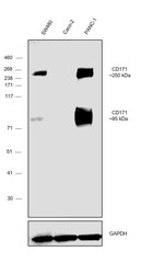
- Western blot was performed using Anti-CD171 Monoclonal Antibody (UJ127) (Product # MA5-14140) and a ~250 and 85 kDa band corresponding to CD171 was observed across cell lines tested except Caco-2 which is reported to be negative. Whole Cell Extract-WCL (30 µg lysate) of SW480 (Lane 1), Caco-2 (Lane 2) and PANC-1 (Lane 3) were electrophoresed using NuPAGE™ 4-12% Bis-Tris Protein Gel (Product # NP0321BOX). Resolved proteins were then transferred onto a Nitrocellulose membrane (Product # IB23001) by iBlot® 2 Dry Blotting System (Product # IB21001). The blot was probed with the primary antibody (1ug/ml) and detected by chemiluminescence with Goat anti-Mouse IgG (H+L) Superclonal™ Recombinant Secondary Antibody, HRP (Product # A28177,1:4000 dilution) using the iBright FL 1000 (Product # A32752). Chemiluminescent detection was performed using Novex® ECL Chemiluminescent Substrate Reagent Kit (Product # WP20005).

Supportive validation
- Submitted by
- Invitrogen Antibodies (provider)
- Main image

- Experimental details
- Immunofluorescence analysis of CD171 was performed using 70% confluent log phase SW480 cells. The cells were fixed with 4% paraformaldehyde for 10 minutes, permeabilized with 0.1% Triton™ X-100 for 15 minutes, and blocked with 2% BSA for 45 minutes at room temperature. The cells were labeled with CD171 Monoclonal Antibody (UJ127) (Product # MA5-14140) at 1:200 dilution in 0.1% BSA, incubated at 4 degree celsius overnight and then labeled with Goat anti-Mouse IgG (H+L) Highly Cross-Adsorbed Secondary Antibody, Alexa Fluor Plus 488 (Product # A32723), (1:3000 dilution), for 45 minutes at room temperature (Panel a: Green). Nuclei (Panel b: Blue) were stained with ProLong™ Diamond Antifade Mountant with DAPI (Product # P36962). F-actin (Panel c: Red) was stained with Rhodamine Phalloidin (Product # R415, 1:300 dilution). Panel d represents the merged image showing membrane localization. Panel e represents merged image for Caco-2 cells showing no staining for CD171. Panel f represents control cells with no primary antibody to assess background. The images were captured at 60X magnification.

Supportive validation
- Submitted by
- Invitrogen Antibodies (provider)
- Main image
- Experimental details
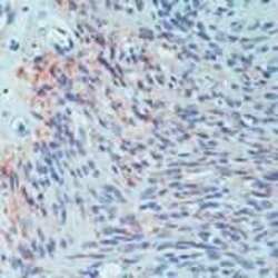
- Formalin-fixed, paraffin-embedded human schwannoma stained with L1 Cell Adhesion Molecule antibody using peroxidase-conjugate and AEC chromogen. Note membrane staining of tumor cells.

- Submitted by
- Invitrogen Antibodies (provider)
- Main image

- Experimental details
- Immunohistochemistry analysis of NCAM1 in 4% PFA, OCT embedded frozen mouse E16.5 lung sections. Sections were blocked and probed with a NCAM1 mouse monoclonal antibody (Product # MA5-14140) at a dilution of 1:1000. Detection was performed using an Alexa Fluor-568-conjugated anti-mouse secondary antibody (Product # A-11004).

Supportive validation
- Submitted by
- Invitrogen Antibodies (provider)
- Main image
- Experimental details
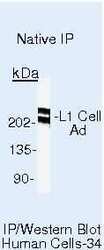
- Immunoprecipitation of L1 Cell Adhesion Molecule using L1 Cell Adhesion Molecule Monoclonal Antibody (Product # MA5-14140) on Native Human IMR-5 Cells.
