 Explore
 Validate
 Learn
Learn Western blot
Antibody data
- Antibody Data
- Antigen structure
- References [1]
- Comments [0]
- Validations
- Western blot [2]
- Immunocytochemistry [1]
Submit
Validation data
Reference
Comment
Report error
- Product number
- AF5104 - Provider product page

- Provider
- R&D Systems
- Product name
- Human LIN-41 Antibody
- Antibody type
- Polyclonal
- Description
- Antigen Affinity-purified. Detects human LIN-41 in direct ELISAs and Western blots.
- Reactivity
- Human
- Host
- Sheep
- Conjugate
- Unconjugated
- Antigen sequence
Q2Q1W2- Isotype
- IgG
- Vial size
- 100 ug
- Concentration
- LYOPH
- Storage
- Use a manual defrost freezer and avoid repeated freeze-thaw cycles. 12 months from date of receipt, -20 to -70 °C as supplied. 1 month, 2 to 8 °C under sterile conditions after reconstitution. 6 months, -20 to -70 °C under sterile conditions after reconstitution.
Submitted references T cell activation induces proteasomal degradation of Argonaute and rapid remodeling of the microRNA repertoire.
Bronevetsky Y, Villarino AV, Eisley CJ, Barbeau R, Barczak AJ, Heinz GA, Kremmer E, Heissmeyer V, McManus MT, Erle DJ, Rao A, Ansel KM
The Journal of experimental medicine 2013 Feb 11;210(2):417-32
The Journal of experimental medicine 2013 Feb 11;210(2):417-32
No comments: Submit comment
Supportive validation
- Submitted by
- R&D Systems (provider)
- Main image

- Experimental details
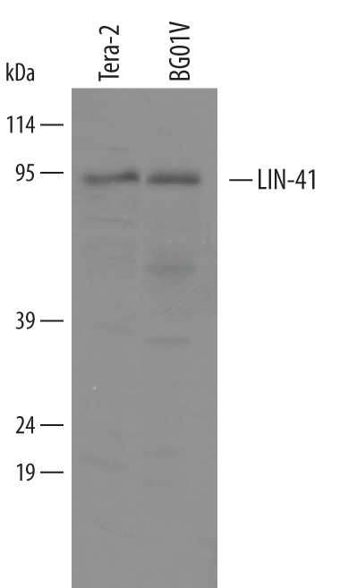
- Detection of Human LIN-41 by Western Blot. Western blot shows lysates of BG0IV human embryonic stem cells and NTera-2 human testicular embryonic carcinoma cell line. PVDF membrane was probed with 1 µg/mL of Sheep Anti-Human LIN-41 Antigen Affinity-purified Polyclonal Antibody (Catalog # AF5104) followed by HRP-conjugated Anti-Sheep IgG Secondary Antibody (Catalog # HAF016). A specific band was detected for LIN-41 at approximately 95 kDa (as indicated). This experiment was conducted under reducing conditions and using Immunoblot Buffer Group 8.
- Submitted by
- R&D Systems (provider)
- Main image
- Experimental details
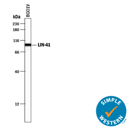
- Detection of Human LIN-41 by Simple WesternTM. Simple Western lane view shows lysates of BG01V human embryonic stem cells, loaded at 0.2 mg/mL. A specific band was detected for LIN-41 at approximately 97 kDa (as indicated) using 10 µg/mL of Sheep Anti-Human LIN-41 Antigen Affinity-purified Polyclonal Antibody (Catalog # AF5104) followed by 1:50 dilution of HRP-conjugated Anti-Sheep IgG Secondary Antibody (Catalog # HAF016). This experiment was conducted under reducing conditions and using the 12-230 kDa separation system.

Supportive validation
- Submitted by
- R&D Systems (provider)
- Main image
- Experimental details
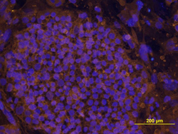
- LIN-41 in BG01V Human Stem Cells. LIN-41 was detected in immersion fixed BG01V human embryonic stem cells using Human LIN-41 Antigen Affinity-purified Polyclonal Antibody (Catalog # AF5104) at 10 µg/mL for 3 hours at room temperature. Cells were stained using the NorthernLights™ 557-conjugated Anti-Sheep IgG Secondary Antibody (yellow; Catalog # NL010) and counterstained with DAPI (blue). View our protocol for Fluorescent ICC Staining of Cells on Coverslips.
