 Explore
 Validate
 Learn
Learn Western blot
Antibody data
- Antibody Data
- Antigen structure
- References [0]
- Comments [0]
- Validations
- Western blot [7]
- Immunocytochemistry [1]
- Immunohistochemistry [1]
Submit
Validation data
Reference
Comment
Report error
- Product number
- PA5-31674 - Provider product page

- Provider
- Invitrogen Antibodies
- Product name
- RAB35 Polyclonal Antibody
- Antibody type
- Polyclonal
- Antigen
- Recombinant protein fragment
- Description
- Recommended positive controls: U87-MG, mouse brain, RAB35-transfected 293T, CHO.
- Concentration
- 0.24 mg/mL
No comments: Submit comment
Supportive validation
- Submitted by
- Invitrogen Antibodies (provider)
- Main image

- Experimental details
- Western blot analysis of RAB35 using 30 µg of U87-MG lysate. Samples were loaded onto a 12% SDS-PAGE gel and probed with a RAB35 polyclonal antibody (Product # PA5-31674) at a dilution of 1:1000.

- Submitted by
- Invitrogen Antibodies (provider)
- Main image

- Experimental details
- Western Blot analysis of RAB35 was performed by separating 30 µg of various whole cell extracts by 12% SDS-PAGE. Proteins were transferred to a membrane and probed with a RAB35 Polyclonal Antibody (Product # PA5-31674) at a dilution of 1:1000 and a HRP-conjugated anti-rabbit IgG secondary antibody.

- Submitted by
- Invitrogen Antibodies (provider)
- Main image

- Experimental details
- Western blot analysis of RAB35 was performed by separating 50 µg of mouse tissue extract by 12% SDS-PAGE. Proteins were transferred to a membrane and probed with a RAB35 Polyclonal Antibody (Product # PA5-31674) at a dilution of 1:1000. The HRP-conjugated anti-rabbit IgG antibody was used to detect the primary antibody.

- Submitted by
- Invitrogen Antibodies (provider)
- Main image

- Experimental details
- Western Blot using RAB35 Polyclonal Antibody (Product # PA5-31674). Mouse tissue extract (50 µg) was separated by 12% SDS-PAGE, and the membrane was blotted with RAB35 Polyclonal Antibody (Product # PA5-31674) diluted at 1:1,000. The HRP-conjugated anti-rabbit IgG antibody was used to detect the primary antibody.

- Submitted by
- Invitrogen Antibodies (provider)
- Main image

- Experimental details
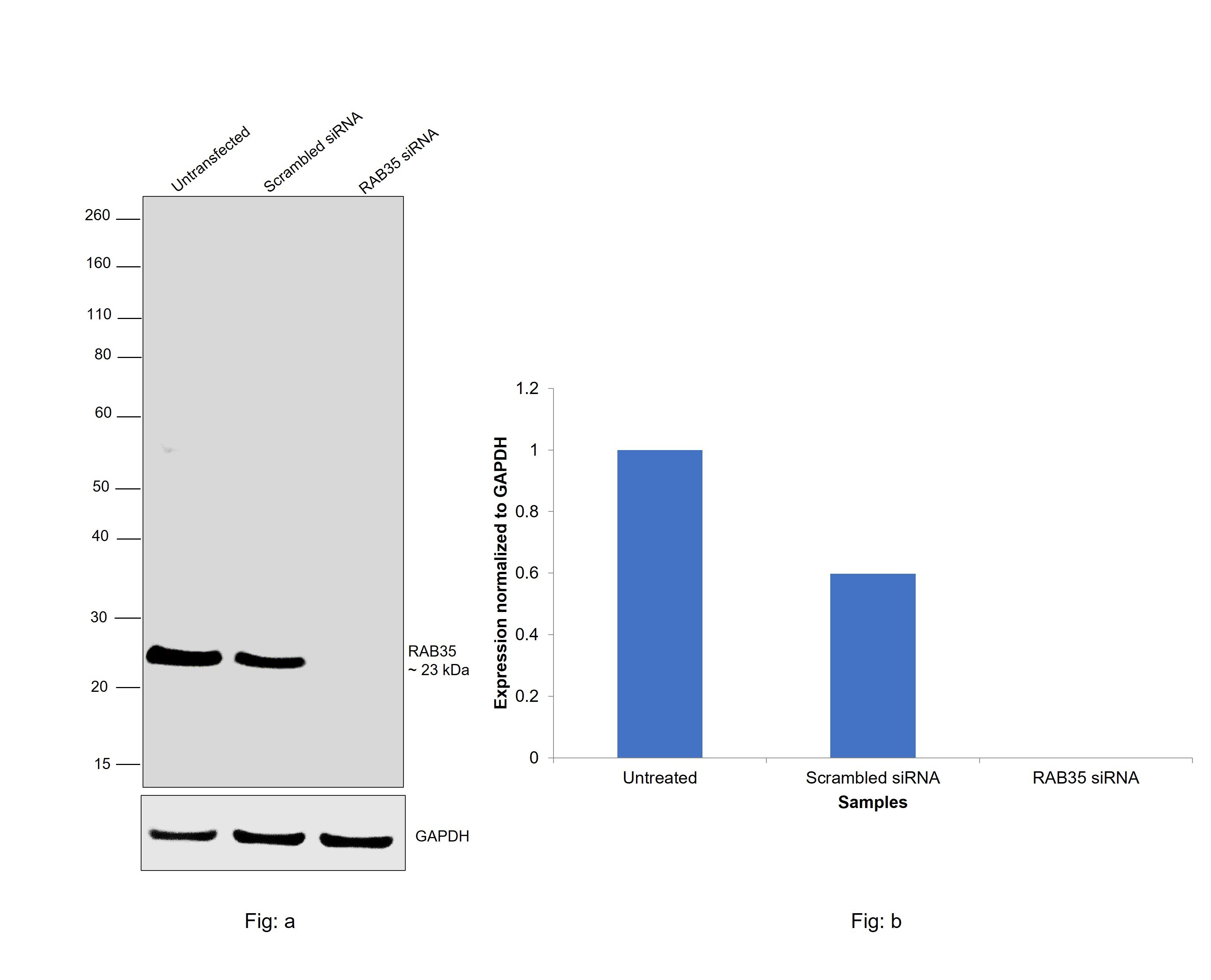
- Knockdown of RAB35 was achieved by transfecting Caki-1 with RAB35 specific siRNAs (Silencer® select Product # SS21707, SS21708). Western blot analysis (Fig. a) was performed using whole cell extracts from the RAB35 knockdown cells (lane 3), non-targeting scrambled siRNA transfected cells (lane 2) and untransfected cells (lane 1). The blot was probed with RAB35 Polyclonal Antibody (Product # PA5-31674, 1:2000) and Goat anti-Rabbit IgG (H+L) Superclonal™ Recombinant Secondary Antibody, HRP (Product # A27036, 1:20,000) and detected by chemiluminescence using the iBright™ FL1500 Imaging System (Product # A44115). Densitometric analysis of this western blot is shown in histogram (Fig. b). Decrease in signal upon siRNA mediated knock down confirms that antibody is specific to RAB35.
- Submitted by
- Invitrogen Antibodies (provider)
- Main image

- Experimental details
- Western Blot analysis of RAB35 was performed by separating 30 µg of non-transfected (–) and transfected (+) 293T whole cell extracts by 12% SDS-PAGE. Proteins were transferred to a membrane and probed with a RAB35 Polyclonal Antibody (Product # PA5-31674) at a dilution of 1:2000. The HRP-conjugated anti-rabbit IgG antibody was used to detect the primary antibody.

- Submitted by
- Invitrogen Antibodies (provider)
- Main image

- Experimental details
- Western blot was performed using RAB35 Polyclonal Antibody (Product # PA5-31674) and a 23 kDa band corresponding to RAB35 was observed across cell lines and tissue tested. Whole cell extracts (30 µg lysate) of Raji (Lane 1), Jurkat (Lane 2), Caki-1 (Lane 3), 769-P (Lane 4), Mouse Cerebellum (Lane 5) and Mouse Lung (Lane 6) were electrophoresed using NuPAGE™ 4-12% Bis-Tris Protein Gel (Product # NP0321BOX), 10 well. Resolved proteins were then transferred onto a nitrocellulose membrane (Product # IB23002) by iBlot® 2 Dry Blotting System (Product # IB21001). The blot was probed with the primary antibody (1:2000) and detected by chemiluminescence with Goat anti-Rabbit IgG (H+L) Superclonal™ Recombinant Secondary Antibody, HRP (Product # A27036, 1:20,000) using the iBright™ FL1500 Imaging System (Product # A44115). Chemiluminescent detection was performed using SuperSignal™ West Atto Ultimate Sensitivity Substrate (Product # A38556).

Supportive validation
- Submitted by
- Invitrogen Antibodies (provider)
- Main image

- Experimental details
- Immunocytochemistry-Immunofluorescence analysis of RAB35 was performed in U87-MG cells fixed in 4% paraformaldehyde at RT for 15 min. Green: RAB35 Polyclonal Antibody (Product # PA5-31674) diluted at 1:500. Blue: Hoechst 33342 staining.

Supportive validation
- Submitted by
- Invitrogen Antibodies (provider)
- Main image

- Experimental details
- Immunohistochemical analysis of paraffin-embedded human breast cancer, using RAB35 (Product # PA5-31674) antibody at 1:250 dilution. Antigen Retrieval: Citrate buffer, pH 6.0, 15 min.
