 Explore
 Validate
 Learn
Learn Western blot
Antibody data
- Antibody Data
- Antigen structure
- References [3]
- Comments [0]
- Validations
- Western blot [1]
- Immunocytochemistry [2]
- Immunohistochemistry [2]
Submit
Validation data
Reference
Comment
Report error
- Product number
- GTX101141 - Provider product page

- Provider
- GeneTex
- Proper citation
- GeneTex Cat#GTX101141, RRID:AB_1240542
- Product name
- N-Cadherin antibody [N1N2], N-term
- Antibody type
- Polyclonal
- Reactivity
- Human, Mouse
- Host
- Rabbit
Submitted references Cross-link regulation of precursor N-cadherin and FGFR1 by GDNF increases U251MG cell viability.
Precursor N-cadherin mediates glial cell line-derived neurotrophic factor-promoted human malignant glioma.
Tumor suppressor SCUBE2 inhibits breast-cancer cell migration and invasion through the reversal of epithelial-mesenchymal transition.
Tang CX, Gu YX, Liu XF, Tong SY, Ayanlaja AA, Gao Y, Ji GQ, Xiong Y, Huang LY, Gao DS
Oncology reports 2018 Jul;40(1):443-453
Oncology reports 2018 Jul;40(1):443-453
Precursor N-cadherin mediates glial cell line-derived neurotrophic factor-promoted human malignant glioma.
Xiong Y, Liu L, Zhu S, Zhang B, Qin Y, Yao R, Zhou H, Gao DS
Oncotarget 2017 Apr 11;8(15):24902-24914
Oncotarget 2017 Apr 11;8(15):24902-24914
Tumor suppressor SCUBE2 inhibits breast-cancer cell migration and invasion through the reversal of epithelial-mesenchymal transition.
Lin YC, Lee YC, Li LH, Cheng CJ, Yang RB
Journal of cell science 2014 Jan 1;127(Pt 1):85-100
Journal of cell science 2014 Jan 1;127(Pt 1):85-100
No comments: Submit comment
Supportive validation
- Submitted by
- GeneTex (provider)
- Main image

- Experimental details
- Sample (30 ug of whole cell lysate) A: K562 5% SDS PAGE GTX101141 diluted at 1:2000

Supportive validation
- Submitted by
- GeneTex (provider)
- Main image

- Experimental details
- N-Cadherin antibody [N1N2], N-term detects N-Cadherin protein at cytoplasm by immunofluorescent analysis.Sample: SH-SY5Y cells were fixed in 4% paraformaldehyde at RT for 15 min.Green: N-Cadherin protein stained by N-Cadherin antibody [N1N2], N-term (GTX101141) diluted at 1:400.Red: beta Tubulin 3/ TUJ1 protein stained by beta Tubulin 3/ TUJ1 antibody (GTX631836) diluted at 1:200.Blue: Hoechst 33342 staining.

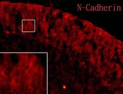
Supportive validation
- Submitted by
- GeneTex (provider)
- Main image
- Experimental details
- N-Cadherin antibody [N1N2] detects N-Cadherin protein on embryonic mouse brain by immunohistochemical analysis. Sample: Frozen section of embryonic mouse brain (mE18.5). Red: N-Cadherin antibody [N1N2](GTX101141) diluted at 1:250.

- Submitted by
- GeneTex (provider)
- Main image

- Experimental details
- N-Cadherin antibody [N1N2] detects N-Cadherin protein on embryonic mouse brain by immunohistochemical analysis. Sample: Frozen section of embryonic mouse brain (mE18.5). N-Cadherin antibody [N1N2] (GTX101141) diluted at 1:500.
