Explore
 Validate
 Learn
Learn Western blot
 Immunoprecipitation
Antibody data
- Antibody Data
- Antigen structure
- References [2]
- Comments [0]
- Validations
- Western blot [1]
- Immunohistochemistry [1]
- Other assay [2]
Submit
Validation data
Reference
Comment
Report error
- Product number
- PA5-17526 - Provider product page

- Provider
- Invitrogen Antibodies
- Product name
- N-cadherin Polyclonal Antibody
- Antibody type
- Polyclonal
- Antigen
- Synthetic peptide
- Description
- It is not recommended to aliquot this antibody.
- Reactivity
- Human, Mouse, Rat, Bovine, Drosophila, Zebrafish
- Host
- Rabbit
- Isotype
- IgG
- Vial size
- 100 µL
- Concentration
- 9 µg/mL
- Storage
- -20°C
Submitted references Blocking Connexin-43 mediated hemichannel activity protects against early tubular injury in experimental chronic kidney disease.
A rare schizophrenia risk variant of CACNA1I disrupts Ca(V)3.3 channel activity.
Price GW, Chadjichristos CE, Kavvadas P, Tang SCW, Yiu WH, Green CR, Potter JA, Siamantouras E, Squires PE, Hills CE
Cell communication and signaling : CCS 2020 May 25;18(1):79
Cell communication and signaling : CCS 2020 May 25;18(1):79
A rare schizophrenia risk variant of CACNA1I disrupts Ca(V)3.3 channel activity.
Andrade A, Hope J, Allen A, Yorgan V, Lipscombe D, Pan JQ
Scientific reports 2016 Oct 19;6:34233
Scientific reports 2016 Oct 19;6:34233
No comments: Submit comment
Supportive validation
- Submitted by
- Invitrogen Antibodies (provider)
- Main image

- Experimental details
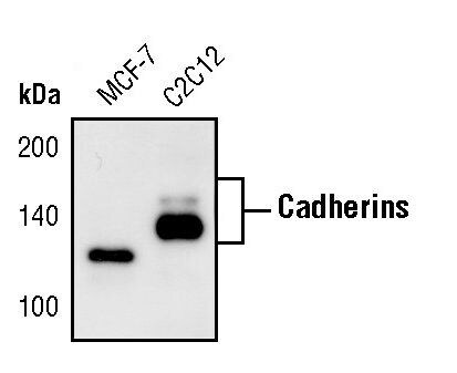
- Western blot analysis of Pan-Cadherin in extracts from MCF-7 and C2C12 cells using Pan-Cadherin polyclonal antibody (Product # PA5-17526).
Supportive validation
- Submitted by
- Invitrogen Antibodies (provider)
- Main image
- Experimental details
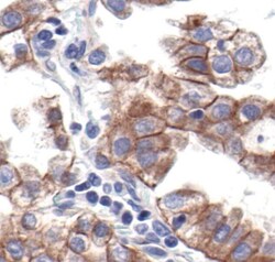
- Immunohistochemical analysis of Pan-Cadherin in paraffin-embedded human bronchioalveolar cell carcinoma using a Pan-Cadherin polyclonal antibody (Product # PA5-17526) showing membrane localization.

Supportive validation
- Submitted by
- Invitrogen Antibodies (provider)
- Main image
- Experimental details
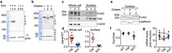
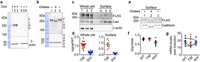
- Figure 2 R1346H affects surface expression of hCa V 3.3. All membranes were cut in two; upper membranes were probed with anti-FLAG to measure hCa V 3.3 levels and lower membranes with control antibodies. ( a ) Anti-FLAG signals from Flp-In T-REx HEK293 cell lysates after induction for 24, 48, and 72 hrs with (lanes 1-3), and without (lanes 4-6), 1 mug/ml doxycycline. ( b ) Anti-FLAG signals in whole cell lysates from cells expressing WT-hCa V 3.3, untreated (1), with glycosidase (2), and same as (2) but lacking glycosidase (3). ( a , b ) Compiled figures from 4 digital images of the same gel. Protein ladder images are juxtaposed to the immunoblots; ladder lane is colored in blue. Dotted lines indicate the spliced borders of two immunoblots. ( c ) Anti-FLAG hCav3.3 levels in whole cell lysate (1-3) and biotin-surface fraction (4-6) from cells expressing WT (1, 4), T797M (T/M) (2, 5), and R1346H (R/H) (3, 6). ( d ) Anti-FLAG signal in whole cell and biotinylated (surface) preparations from cells expressing T/M and R/H shown relative to WT and normalized to controls (cadherin and beta-actin). Mean +- SE values for T/M were 1.12 +- 0.08 (n = 18, whole cell) and 0.87 +- 0.13 (n = 7, surface); for R/H were 0.29 +- 0.03 (n = 18, whole cell) and 0.12 +- 0.03 (n = 7, surface). Coefficient of variation: T/M, 31% and R/H, 40% (1000 samples bootstrapping). ( e ) Anti-FLAG signals in biotinylated surface protein from cells expressing WT-hCa V 3.3, untreated (1), glycosidase exposure (2) a
- Submitted by
- Invitrogen Antibodies (provider)
- Main image

- Experimental details
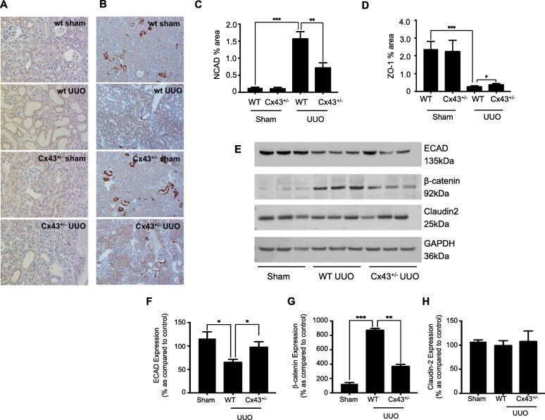
- Fig. 8 Cx43 +/- mice exhibit minimal disassembly of the adherens and tight junction complex. Quantification of immunohistochemical staining determined an increase in N-cadherin ( a & c ) and decrease in ZO-1 ( b & d ) expression in wildtype (WT) UUO compared to WT sham controls. In Cx43 +/- (UUO) mice, expression of both markers was partially restored to near basal levels. Furthermore, western blotting ( e ) of renal cortex determined changes in expression of E-cadherin ( f ), beta-catenin ( g ) and Claudin-2 ( h ) in WT UUO mice compared to WT shams, congruent to those found in vitro when cells were treated with TGF-beta1. As expected, these changes were partially negated in Cx43 +/- (UUO) mice. Results were from six separate experiments; with significance shown: * P < 0.05; ** P < 0.01; *** P < 0.001