Explore
 Validate
 Learn
Learn Western blot
Antibody data
- Antibody Data
- Antigen structure
- References [4]
- Comments [0]
- Validations
- Western blot [9]
- Immunocytochemistry [1]
- Immunohistochemistry [3]
- Other assay [3]
Submit
Validation data
Reference
Comment
Report error
- Product number
- PA5-29570 - Provider product page

- Provider
- Invitrogen Antibodies
- Product name
- N-cadherin Polyclonal Antibody
- Antibody type
- Polyclonal
- Antigen
- Recombinant protein fragment
- Description
- Recommended positive controls: NT2D1, U87-MG, SK-N-SH, IMR-32, SK-N-AS, mouse brain, rat brain, PC-12, Rat2.
- Concentration
- 0.19 mg/mL
Submitted references Poria Acid, Triterpenoids Extracted from Poria cocos, Inhibits the Invasion and Metastasis of Gastric Cancer Cells.
Betulonic Acid, as One of the Active Components of the Celastrus orbiculatus Extract, Inhibits the Invasion and Metastasis of Gastric Cancer Cells by Mediating Cytoskeleton Rearrangement In Vitro.
A truncating mutation in the autophagy gene UVRAG drives inflammation and tumorigenesis in mice.
Truncating mutation in the autophagy gene UVRAG confers oncogenic properties and chemosensitivity in colorectal cancers.
Wang H, Luo Y, Chu Z, Ni T, Ou S, Dai X, Zhang X, Liu Y
Molecules (Basel, Switzerland) 2022 Jun 6;27(11)
Molecules (Basel, Switzerland) 2022 Jun 6;27(11)
Betulonic Acid, as One of the Active Components of the Celastrus orbiculatus Extract, Inhibits the Invasion and Metastasis of Gastric Cancer Cells by Mediating Cytoskeleton Rearrangement In Vitro.
Chu Z, Luo Y, Ni T, Zhu M, Feng X, Liu Y, Wang H
Molecules (Basel, Switzerland) 2022 Feb 2;27(3)
Molecules (Basel, Switzerland) 2022 Feb 2;27(3)
A truncating mutation in the autophagy gene UVRAG drives inflammation and tumorigenesis in mice.
Quach C, Song Y, Guo H, Li S, Maazi H, Fung M, Sands N, O'Connell D, Restrepo-Vassalli S, Chai B, Nemecio D, Punj V, Akbari O, Idos GE, Mumenthaler SM, Wu N, Martin SE, Hagiya A, Hicks J, Cui H, Liang C
Nature communications 2019 Dec 12;10(1):5681
Nature communications 2019 Dec 12;10(1):5681
Truncating mutation in the autophagy gene UVRAG confers oncogenic properties and chemosensitivity in colorectal cancers.
He S, Zhao Z, Yang Y, O'Connell D, Zhang X, Oh S, Ma B, Lee JH, Zhang T, Varghese B, Yip J, Dolatshahi Pirooz S, Li M, Zhang Y, Li GM, Ellen Martin S, Machida K, Liang C
Nature communications 2015 Aug 3;6:7839
Nature communications 2015 Aug 3;6:7839
No comments: Submit comment
Supportive validation
- Submitted by
- Invitrogen Antibodies (provider)
- Main image

- Experimental details
- Western blot analysis of N-cadherin/CDH2 using 30 µg of K562 lysate. Samples were loaded onto a 7.5% SDS-PAGE gel and probed with a N-cadherin/CDH2 polyclonal antibody (Product # PA5-29570) at a dilution of 1:1000.
- Submitted by
- Invitrogen Antibodies (provider)
- Main image

- Experimental details
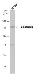
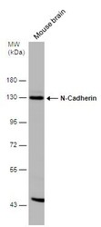
- Western blot analysis of N-cadherin/CDH2 using 50 µg of mouse brain lysate. Samples were loaded onto a 7.5% SDS-PAGE gel and probed with a N-cadherin/CDH2 polyclonal antibody (Product # PA5-29570) at a dilution of 1:1000.

- Submitted by
- Invitrogen Antibodies (provider)
- Main image

- Experimental details
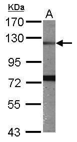
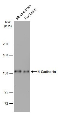
- Western Blot analysis of N-cadherin was performed by separating 50 µg of Rat tissue extracts by 7.5% SDS-PAGE. Proteins were transferred to a membrane and probed with a N-cadherin Polyclonal Antibody (Product # PA5-29570) at a dilution of 1:1000. The HRP-conjugated anti-rabbit IgG antibody was used to detect the primary antibody.

- Submitted by
- Invitrogen Antibodies (provider)
- Main image

- Experimental details
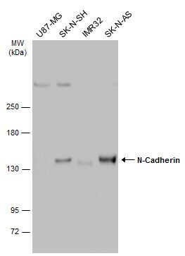
- Western Blot using N-cadherin Polyclonal Antibody (Product # PA5-29570). Various whole cell extracts (30 µg) were separated by 5% SDS-PAGE, and the membrane was blotted with N-cadherin Polyclonal Antibody (Product # PA5-29570) diluted at 1:1,000. The HRP-conjugated anti-rabbit IgG antibody was used to detect the primary antibody.
- Submitted by
- Invitrogen Antibodies (provider)
- Main image

- Experimental details
- N-cadherin Polyclonal Antibody detects N-Cadherin protein by western blot analysis. A. 30 µg PC-12 whole cell extract. B. 30 µg Rat2 whole cell extract.5% SDS-PAGE. N-cadherin Polyclonal Antibody (Product # PA5-29570) dilution: 1:1,000. The HRP-conjugated anti-rabbit IgG antibody was used to detect the primary antibody.

- Submitted by
- Invitrogen Antibodies (provider)
- Main image
- Experimental details
- Western Blot analysis of N-cadherin was performed by separating 30 µg of Whole cell extracts by 7.5% SDS-PAGE. Proteins were transferred to a membrane and probed with a N-cadherin Polyclonal Antibody (Product # PA5-29570) at a dilution of 1:1000. The HRP-conjugated anti-rabbit IgG antibody was used to detect the primary antibody.

- Submitted by
- Invitrogen Antibodies (provider)
- Main image

- Experimental details
- Western Blot using N-cadherin Polyclonal Antibody (Product # PA5-29570). Various tissue extracts (50 µg) were separated by 5% SDS-PAGE, and the membrane was blotted with N-cadherin Polyclonal Antibody (Product # PA5-29570) diluted at 1:1,000. The HRP-conjugated anti-rabbit IgG antibody was used to detect the primary antibody.
- Submitted by
- Invitrogen Antibodies (provider)
- Main image
- Experimental details
- Western Blot analysis of N-cadherin was performed by separating 50 µg of Mouse tissue extracts by 7.5% SDS-PAGE. Proteins were transferred to a membrane and probed with a N-cadherin Polyclonal Antibody (Product # PA5-29570) at a dilution of 1:500. The HRP-conjugated anti-rabbit IgG antibody was used to detect the primary antibody.

- Submitted by
- Invitrogen Antibodies (provider)
- Main image
- Experimental details
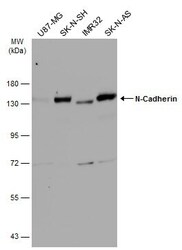
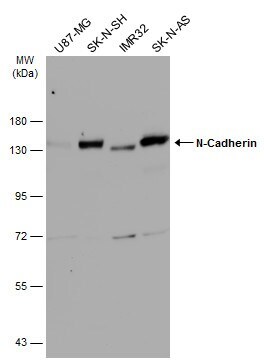
- Western Blot analysis of N-cadherin was performed by separating 30 µg of various whole cell extracts by 7.5% SDS-PAGE. Proteins were transferred to a membrane and probed with a N-cadherin Polyclonal Antibody (Product # PA5-29570) at a dilution of 1:1000 and a HRP-conjugated anti-rabbit IgG secondary antibody.
Supportive validation
- Submitted by
- Invitrogen Antibodies (provider)
- Main image
- Experimental details
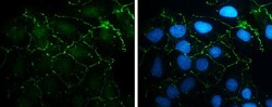
- Immunocytochemistry-Immunofluorescence analysis of N-cadherin was performed in NT2D1 cells fixed in 4% paraformaldehyde at RT for 15 min. Green: N-cadherin Polyclonal Antibody (Product # PA5-29570) diluted at 1:500. Blue: Hoechst 33342 staining.

Supportive validation
- Submitted by
- Invitrogen Antibodies (provider)
- Main image
- Experimental details
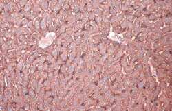
- Immunohistochemistry (Paraffin) analysis of N-cadherin was performed in paraffin-embedded mouse liver tissue using N-cadherin Polyclonal Antibody (Product # PA5-29570) at a dilution of 1:500. Antigen Retrieval: Citrate buffer, pH 6.0, 15 min.

- Submitted by
- Invitrogen Antibodies (provider)
- Main image
- Experimental details
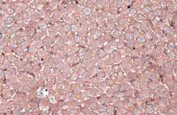
- Immunohistochemistry (Paraffin) analysis of N-cadherin was performed in paraffin-embedded rat liver tissue using N-cadherin Polyclonal Antibody (Product # PA5-29570) at a dilution of 1:500. Antigen Retrieval: Citrate buffer, pH 6.0, 15 min.

- Submitted by
- Invitrogen Antibodies (provider)
- Main image
- Experimental details
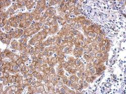
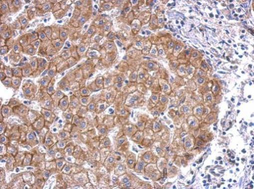
- N-cadherin Polyclonal Antibody detects CDH2 protein at membrane on hepatoma by immunohistochemical analysis. Sample: Paraffin-embedded human hepatoma. N-cadherin Polyclonal Antibody (Product # PA5-29570) dilution: 1:500. Antigen Retrieval: EDTA based buffer, pH 8.0, 15 min.
Supportive validation
- Submitted by
- Invitrogen Antibodies (provider)
- Main image
- Experimental details
- Figure 5 UVRAG FS activates Rac-1 and promotes tumour metastasis in vitro and vivo . ( a ) Rac-1 activation by UVRAG FS . Western blot shows a pull-down experiment to detect GTP-bound Rac1 in SW480.UVRAG FS cells and on drug treatment. Histogram shows quantification from three independent experiments. ( b ) Representative images of scratch-wound healing exhibit the motility of SW480.UVRAG FS cells. Cell motility into the wound area was taken at 0 and 16 h as marked by red lines. Wound-healing index was quantified (right). Data are the means+-s.d. ( n =3). ** P

- Submitted by
- Invitrogen Antibodies (provider)
- Main image

- Experimental details
- The effects of PA on the expression of EMT and MMPs. ( A , C - E ) The Western blot band and quantification relative statistics of EMT-related proteins in the AGS cells. ( B , F - H ) The Western blot band and quantification relative statistics of the metastasis-associated proteins in AGS cells. * p < 0.05, ** p < 0.01, *** p < 0.001, **** p < 0.0001.

- Submitted by
- Invitrogen Antibodies (provider)
- Main image

- Experimental details
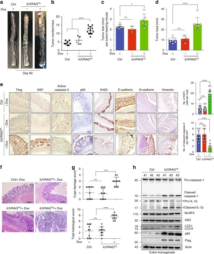
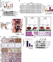
- Fig. 5 UVRAG FS predisposes mice to colitis-associated colon cancer. a Representative macroscopic images of the colon from AOM-DSS-treated control (Ctrl) and iUVRAG FS mice in the presence/absence of Dox at the time of necropsy (day 60). Red arrows highlight colonic tumors. b - d The number ( b ), size ( c ), and overall load ( d ) of colonic tumors in AOM-DSS-treated mice in a . Tumor load is evaluated by totaling the diameters of all tumors in AOM-DSS-treated mice in a . n = 5-12 mice per genotype. e IHC staining of Ki67, cleaved caspase 3, p62, keratin 20, E-cadherin, N-cadherin, and vimentin in the colons from AOM-DSS-treated mice of indicated genotypes. Data are from one animal that is representative of 5-12 animals in each group. The levels of Ki67 staining (top right) and tumor apoptosis (bottom right) in the indicated colon were quantified. Arrows (red) denote cells undergoing apoptosis. Scale bars, 100 mum. f H&E-stained sections of the colon from AOM-DSS-treated mice in a . Data are from one animal that is representative of 5-12 animals in each group. Scale bars, 100 mum. g Histological scores for crypt damage (top) and total histological score (bottom) of colons from mice in a were quantified at day 60 as described in the Methods. n = 10 mice per group. h WB analysis of caspase-1 cleavage, IL-1beta production, LC3-I/II, and p62 levels in colon tissues of Dox-treated control and iUVRAG FS mice after AOM-DSS treatment. Data in h are from one experiment that is repres