 Explore
 Validate
 Learn
Learn Western blot
Antibody data
- Antibody Data
- Antigen structure
- References [1]
- Comments [0]
- Validations
- Western blot [2]
- Immunohistochemistry [2]
- Other assay [1]
Submit
Validation data
Reference
Comment
Report error
- Product number
- PA5-20864 - Provider product page

- Provider
- Invitrogen Antibodies
- Product name
- PPAPDC1A Polyclonal Antibody
- Antibody type
- Polyclonal
- Antigen
- Synthetic peptide
- Description
- A suggested positive control is human brain tissue lysate.
- Concentration
- 1 mg/mL
Submitted references Extracellular Vesicles Carry lncRNA SNHG16 to Promote Metastasis of Breast Cancer Cells via the miR-892b/PPAPDC1A Axis.
Xia W, Liu Y, Cheng T, Xu T, Dong M, Hu X
Frontiers in cell and developmental biology 2021;9:628573
Frontiers in cell and developmental biology 2021;9:628573
No comments: Submit comment
Supportive validation
- Submitted by
- Invitrogen Antibodies (provider)
- Main image

- Experimental details
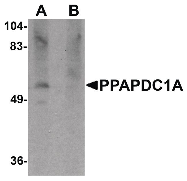
- Western blot analysis of human brain tissue lysate using a PPAPDC1A polyclonal antibody (Product # PA5-20864) at 1 µg/mL in either (A) the presence and (B) the absence of blocking peptide.
- Submitted by
- Invitrogen Antibodies (provider)
- Main image

- Experimental details
- Western Blot analysis of PPAPDC1A in human brain tissue lysate with PPAPDC1A Polyclonal Antibody (Product # PA5-20864) at 1 µg/mL in (A) the absence and (B) the presence of blocking peptide.

Supportive validation
- Submitted by
- Invitrogen Antibodies (provider)
- Main image
- Experimental details
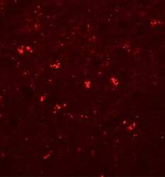
- Immunofluorescence of PPAPDC1A in human brain tissue with PPAPDC1A Polyclonal Antibody (Product # PA5-20864) at 20 µg/mL.
- Submitted by
- Invitrogen Antibodies (provider)
- Main image

- Experimental details
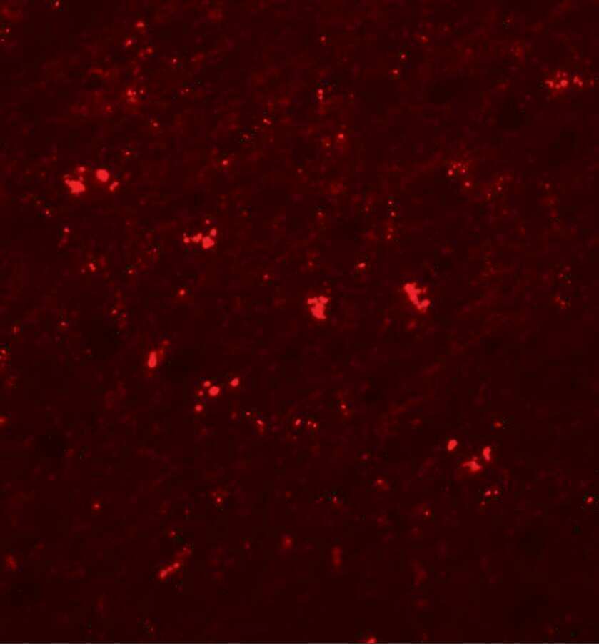
- Immunofluorescence of PPAPDC1A in human brain tissue with PPAPDC1A Polyclonal Antibody (Product # PA5-20864) at 20 µg/mL.
Supportive validation
- Submitted by
- Invitrogen Antibodies (provider)
- Main image

- Experimental details
- FIGURE 6 SNHG16 carried by EVs promoted migration and invasion BC cells by inhibiting miR-892b and upregulating PPAPDC1A. MDA-MB-157 and HS578T cells were transfected with miR-892b-mimic or si-PPAPDC1A, followed by MDA-MB-231 cell-EVs treatment. (A) The miR-892b and PPAPDC1A expression in MDA-MB-157 and HS578T cells was detected using RT-qPCR. (B) The PPAPDC1A protein level in MDA-MB-157 and HS578T cells was detected using Western blotting. (C,D) The migration and invasion of MDA-MB-157 and HS578T cells were measured using Transwell assays. MDA-MB-157 cells and HS578T cells were treated with pcDNA3.1-SNHG16 and miR-892b-mimic. (E) PPAPDC1A mRNA expression in cells was detected using RT-qPCR. (F) PPAPDC1A protein level in cells was detected using Western blotting. Cell experiment was repeated three times. Data were presented as mean +- standard deviation. Data in A and B were analyzed using one-way ANOVA, and data in C and D were analyzed using two-way ANOVA, followed by Tukey's multiple comparison test, * p < 0.05 vs. EVs + mimic-NC group, # p < 0.05 vs. EVs + si-NC group; @ p < 0.05.
