Explore
 Validate
 Learn
Learn Western blot
Antibody data
- Antibody Data
- Antigen structure
- References [0]
- Comments [0]
- Validations
- Western blot [2]
- Immunocytochemistry [1]
Submit
Validation data
Reference
Comment
Report error
- Product number
- PA5-48099 - Provider product page

- Provider
- Invitrogen Antibodies
- Product name
- TEX19 Polyclonal Antibody
- Antibody type
- Polyclonal
- Antigen
- Recombinant full-length protein
- Description
- In direct ELISAs, less than 2% cross-reactivity with recombinant human TEX11 is observed.
- Concentration
- 0.2 mg/mL
No comments: Submit comment
Supportive validation
- Submitted by
- Invitrogen Antibodies (provider)
- Main image
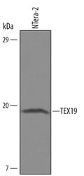
- Experimental details
- Western blot analysis from lysates of NTera-2 human testicular embryonic carcinoma cell line. PVDF Membrane was probed with 0.5 µg/mL of human TEX19 Antigen Affinity-purified Polyclonal Antibody (Product # PA5-48099) followed by HRP-conjugated Anti-Sheep IgG Secondary Antibody. A specific band was detected for TEX19 at approximately 18 kDa (as indicated). This experiment was conducted under reducing conditions.
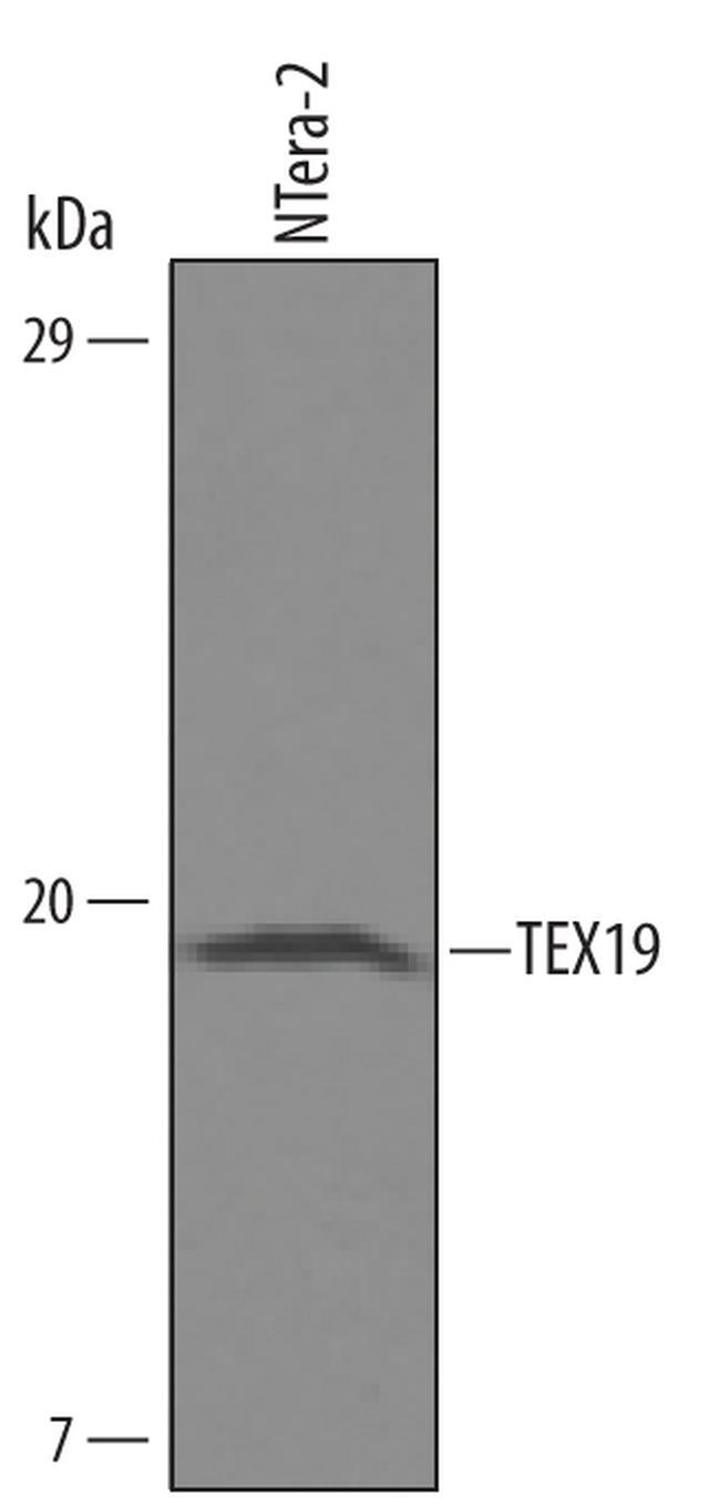
- Submitted by
- Invitrogen Antibodies (provider)
- Main image

- Experimental details
- Western blot analysis of TEX19 in NTera‚2 human testicular embryonic carcinoma cell line. Samples were incubated in TEX19 polyclonal antibody (Product # PA5-48099) using a dilution of 0.5 µg/mL followed by a HRP-conjugated Anti-Sheep IgG secondary antibody. A specific band was detected for TEX19 at approximately 18 kDa (as indicated). This experiment was conducted under reducing conditions.

Supportive validation
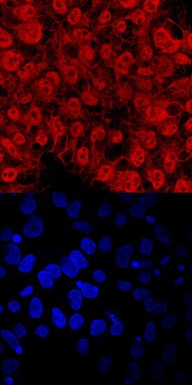
- Submitted by
- Invitrogen Antibodies (provider)
- Main image

- Experimental details
- Immunocytochemistry analysis of TEX19 in immersion fixed BG01V human embryonic stem cells. Samples were incubated in TEX19 polyclonal antibody (Product # PA5-48099) using a dilution of 10 µg/mL for 3 hours at room temperature followed by NorthernLights™ 557-conjugated Anti-Sheep IgG Secondary Antibody (red, upper panel) and counterstained with DAPI (blue, lower panel). Specific staining was localized to nuclei and cytoplasm.