Explore
 Validate
 Learn
Learn Western blot
 Immunoprecipitation
Antibody data
- Antibody Data
- Antigen structure
- References [0]
- Comments [0]
- Validations
- Western blot [3]
- Immunocytochemistry [1]
- Immunohistochemistry [5]
- Other assay [1]
Submit
Validation data
Reference
Comment
Report error
- Product number
- MA5-27531 - Provider product page

- Provider
- Invitrogen Antibodies
- Product name
- CHCHD10 Monoclonal Antibody (OTI4C12)
- Antibody type
- Monoclonal
- Antigen
- Recombinant protein fragment
- Reactivity
- Human
- Host
- Mouse
- Isotype
- IgG
- Antibody clone number
- OTI4C12
- Vial size
- 100 µL
- Concentration
- 1 mg/mL
- Storage
- -20° C, Avoid Freeze/Thaw Cycles
No comments: Submit comment
Supportive validation
- Submitted by
- Invitrogen Antibodies (provider)
- Main image
- Experimental details
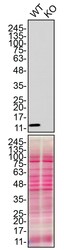
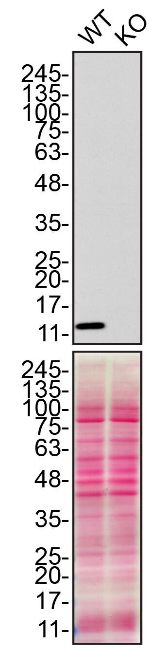
- Western blot of CHCHD10 was performed by loading 50 µg of WT (lane 1) and CHCHD10 CRISPR KO (lane 2) HAP1 cell lysates in RIPA buffer onto a 8-16% gradient polyacrylamide gel. Proteins on the blots were visualized with Ponceau staining (below immunoblot). Proteins were transferred to nitrocellulose membrane and blocked in 5% milk for 1 hr. CHCHD10 was detected at approximately 14 kDa using a CHCHD10 monoclonal antibody (Product # MA5-27531) at a dilution of 1:500 in 5% BSA in TBS with 0.1% Tween 20 (TBST) overnight at 4°C. The peroxidase-conjugated secondary antibody (Product # 62-6520) was diluted to 0.2 µg/mL in TBST with 5% milk for 1 hr. Chemiluminescent detection was performed using Pierce ECL Western Blotting Substrate (Product # 32106). Data courtesy of YCharOS Inc., an open science company with the mission of characterizing commercially available antibodies using knockout validation.
- Submitted by
- Invitrogen Antibodies (provider)
- Main image
- Experimental details
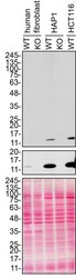
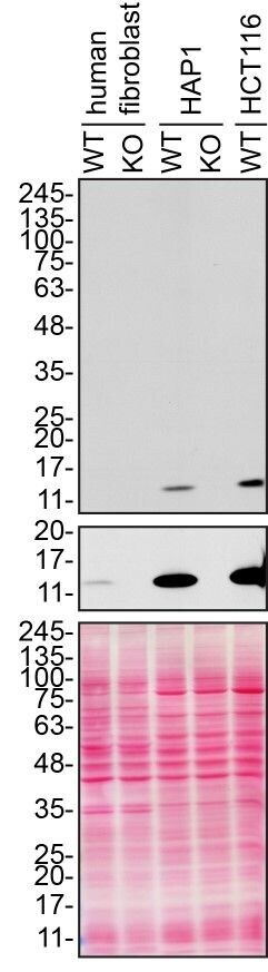
- Western blot of CHCHD10 was performed by loading 80 µg of human fibroblast (WT and CHCHD10 KO), HAP1 (WT and CHCHD10 KO), and HCT116 (WT) lysates in RIPA buffer onto a 8-16% gradient polyacrylamide gel. Proteins on the blots were visualized with Ponceau staining (below immunoblot). Proteins were transferred to nitrocellulose membrane and blocked in 5% milk for 1 hr. CHCHD10 was detected at approximately 14 kDa using a CHCHD10 monoclonal antibody (Product # MA5-27531) at a dilution of 1:500 in 5% BSA in TBS with 0.1% Tween 20 (TBST) overnight at 4°C. The peroxidase-conjugated secondary antibody (Product # 62-6520) was diluted to 0.2 µg/mL in TBST with 5% milk for 1 hr. Chemiluminescent detection was performed using Pierce ECL Western Blotting Substrate (Product # 32106). Data courtesy of YCharOS Inc., an open science company with the mission of characterizing commercially available antibodies using knockout validation.
- Submitted by
- Invitrogen Antibodies (provider)
- Main image
- Experimental details
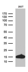
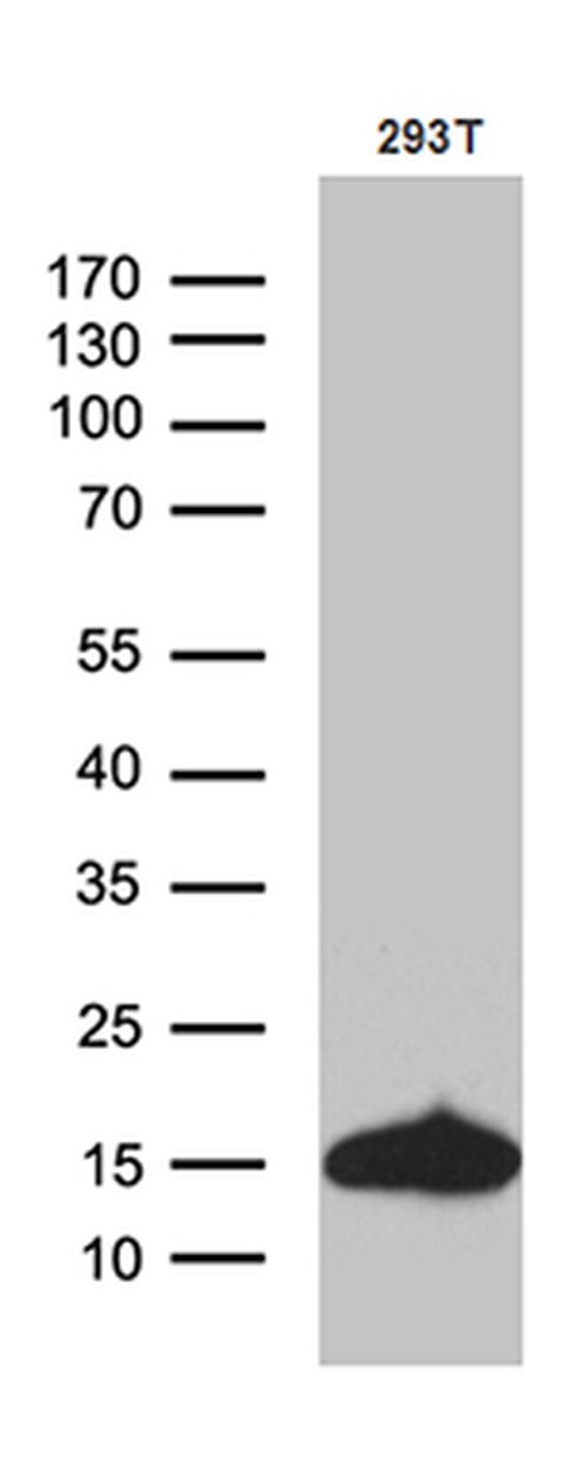
- Western blot analysis of CHCHD10 in H293T cells using 35 µg per lane. Samples were separated by SDS-PAGE and probed with CHCHD10 (Product # MA5-27531) monoclonal antibody at a dilution of 1:500.
Supportive validation
- Submitted by
- Invitrogen Antibodies (provider)
- Main image
- Experimental details
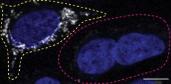
- Immunofluorescence of CHCHD10 was performed using HCT116 wild-type and CHCHD10 KO cells that were transfected with a green or a deep red fluorescent dye, respectively. Post-transfection, WT and KO cells were mixed and plated to a 1:1 ratio on coverslips as a mosaic and incubated for 24 hrs. Cells were fixed in 4% PFA (in PBS) for 15 min; cells were permeabilized with 0.1% Triton X-100 for 10 min at RT and blocked with PBS with 5% BSA, 5% goat serum, and 0.01% Triton X-100 for 30 min. Cells were stained with the CHCHD10 monoclonal antibody (Product # MA5-27531) at a 1:100 dilution overnight at 4°C. Secondary antibody incubation was performed using 1 µg/mL of Goat anti-Mouse IgG (H+L) Highly Cross-Adsorbed Secondary Antibody, Alexa Fluor 555 antibody (Product # A21424) together with DAPI for 1 hr. Imaging was performed with a 40X oil objective and analysis was performed using Image J. Cell image represents a single focal plane; WT and KO cells are outlined with a yellow (WT) or magenta (KO) dashed line. Data courtesy of YCharOS Inc., an open science company with the mission of characterizing commercially available antibodies using knockout validation.

Supportive validation
- Submitted by
- Invitrogen Antibodies (provider)
- Main image
- Experimental details
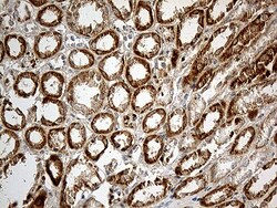
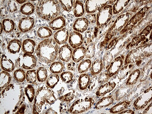
- Immunohistochemistry was performed on paraffin-embedded human kidney tissue. To expose target proteins, heat-induced epitope retrieval by 10mM citric buffer, pH6.0, 100°C for 10min. Following antigen retrieval, tissues were probed with a CHCHD10 monoclonal antibody (Product # MA5-27531) at a dilution of 1:500.
- Submitted by
- Invitrogen Antibodies (provider)
- Main image
- Experimental details
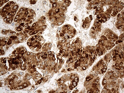
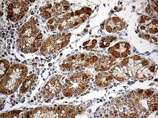
- Immunohistochemistry was performed on paraffin-embedded human liver tissue. To expose target proteins, heat-induced epitope retrieval by 10mM citric buffer, pH6.0, 100°C for 10min. Following antigen retrieval, tissues were probed with a CHCHD10 monoclonal antibody (Product # MA5-27531) at a dilution of 1:500.

- Submitted by
- Invitrogen Antibodies (provider)
- Main image

- Experimental details
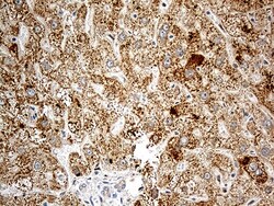
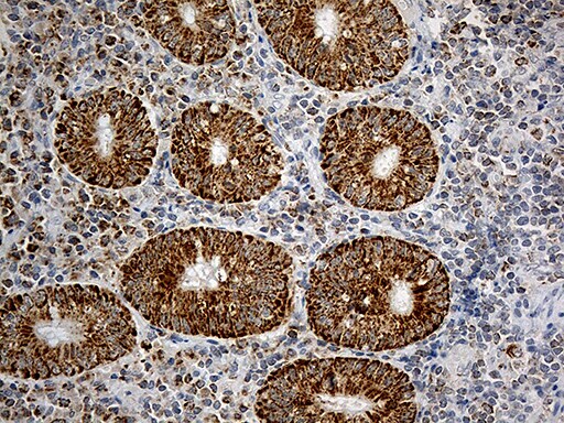
- Immunohistochemistry was performed on paraffin-embedded carcinoma of human liver tissue. To expose target proteins, heat-induced epitope retrieval by 10mM citric buffer, pH6.0, 100°C for 10min. Following antigen retrieval, tissues were probed with a CHCHD10 monoclonal antibody (Product # MA5-27531) at a dilution of 1:500.
- Submitted by
- Invitrogen Antibodies (provider)
- Main image
- Experimental details
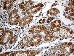
- Immunohistochemistry was performed on paraffin-embedded human gastric tissue. To expose target proteins, heat-induced epitope retrieval by 10mM citric buffer, pH6.0, 100°C for 10min. Following antigen retrieval, tissues were probed with a CHCHD10 monoclonal antibody (Product # MA5-27531) at a dilution of 1:500.
- Submitted by
- Invitrogen Antibodies (provider)
- Main image

- Experimental details
- Immunohistochemistry was performed on paraffin-embedded human appendix tissue. To expose target proteins, heat-induced epitope retrieval by 10mM citric buffer, pH6.0, 100°C for 10min. Following antigen retrieval, tissues were probed with a CHCHD10 monoclonal antibody (Product # MA5-27531) at a dilution of 1:500.
Supportive validation
- Submitted by
- Invitrogen Antibodies (provider)
- Main image
- Experimental details
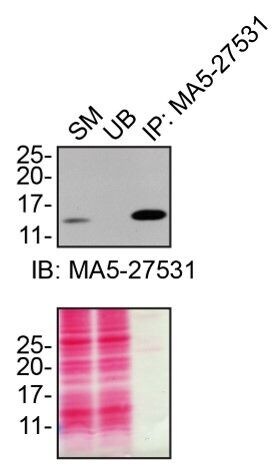
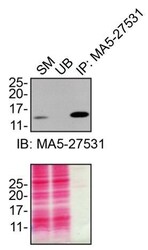
- Immunoprecipitation of CHCHD10 was performed on HCT116 cell lysates. Antibody-bead conjugates were prepared by adding 1 µg of CHCHD10 monoclonal antibody (Product # MA5-27531) with 30 µL of protein G-Sepharose beads and rocked overnight at 4°C. 1 mg of lysate was incubated with an antibody-bead conjugate for 2 hours at 4°C. Following centrifugation and multiple washes, 10% starting material (SM), 10% unbound fraction (UB) and immunoprecipitated fraction (IP) were processed for immunoblot using the same antibody. Ponceau stained transfer of blot is shown. Data courtesy of YCharOS Inc., an open science company with the mission of characterizing commercially available antibodies using knockout validation.