 Explore
 Validate
 Learn
Learn Western blot
Antibody data
- Antibody Data
- Antigen structure
- References [4]
- Comments [0]
- Validations
- Western blot [2]
- Immunocytochemistry [1]
- Other assay [5]
Submit
Validation data
Reference
Comment
Report error
- Product number
- 433270 - Provider product page

- Provider
- Invitrogen Antibodies
- Product name
- Pannexin 3 Polyclonal Antibody
- Antibody type
- Polyclonal
- Antigen
- Synthetic peptide
- Reactivity
- Human, Mouse, Rat
- Host
- Rabbit
- Isotype
- IgG
- Vial size
- 100 µg
- Concentration
- 0.25 mg/mL
- Storage
- Maintain refrigerated at 2-8°C for up to 1 month. For long term storage store at -20°C
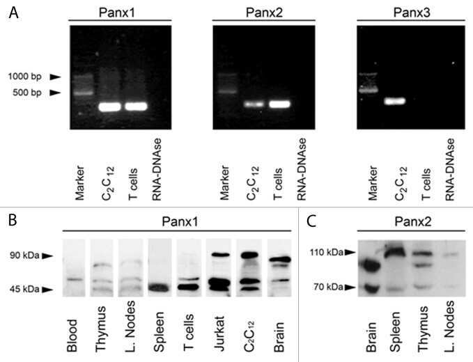
Submitted references Human stem cells express pannexins.
Influence of the three-dimensional culture of human bone marrow mesenchymal stromal cells within a macroporous polysaccharides scaffold on Pannexin 1 and Pannexin 3.
Pannexin1 channels act downstream of P2X 7 receptors in ATP-induced murine T-cell death.
Hyposmotic stress causes ATP release and stimulates Na,K-ATPase activity in porcine lens.
Hainz N, Beckmann A, Schubert M, Haase A, Martin U, Tschernig T, Meier C
BMC research notes 2018 Jan 22;11(1):54
BMC research notes 2018 Jan 22;11(1):54
Influence of the three-dimensional culture of human bone marrow mesenchymal stromal cells within a macroporous polysaccharides scaffold on Pannexin 1 and Pannexin 3.
Guerrero J, Oliveira H, Aid R, Bareille R, Siadous R, Letourneur D, Mao Y, Kohn J, Amédée J
Journal of tissue engineering and regenerative medicine 2018 Apr;12(4):e1936-e1949
Journal of tissue engineering and regenerative medicine 2018 Apr;12(4):e1936-e1949
Pannexin1 channels act downstream of P2X 7 receptors in ATP-induced murine T-cell death.
Shoji KF, Sáez PJ, Harcha PA, Aguila HL, Sáez JC
Channels (Austin, Tex.) 2014;8(2):142-56
Channels (Austin, Tex.) 2014;8(2):142-56
Hyposmotic stress causes ATP release and stimulates Na,K-ATPase activity in porcine lens.
Shahidullah M, Mandal A, Beimgraben C, Delamere NA
Journal of cellular physiology 2012 Apr;227(4):1428-37
Journal of cellular physiology 2012 Apr;227(4):1428-37
No comments: Submit comment
Supportive validation
- Submitted by
- Invitrogen Antibodies (provider)
- Main image

- Experimental details
- Western blot analysis of non-transfected C6 cells (lane B) or cells transfected with rat Pannexin 3 cDNA using Rabbit anti-Pannexin 3 antibody (Product # 433270).

- Submitted by
- Invitrogen Antibodies (provider)
- Main image

- Experimental details
- Western blot analysis of non-transfected C6 cells (lane B) or cells transfected with rat Pannexin 3 cDNA using Rabbit anti-Pannexin 3 antibody (Product # 433270).

Supportive validation
- Submitted by
- Invitrogen Antibodies (provider)
- Main image

- Experimental details
- Immunofluorescence of non-transfected HeLa cells (panel A) and cells transfected with rat Pannexin 3 cDNA using Rabbit anti-Pannexin 3 antibody (Product # 433270).

Supportive validation
- Submitted by
- Invitrogen Antibodies (provider)
- Main image

- Experimental details
- NULL

- Submitted by
- Invitrogen Antibodies (provider)
- Main image

- Experimental details
- NULL
- Submitted by
- Invitrogen Antibodies (provider)
- Main image
- Experimental details
- NULL

- Submitted by
- Invitrogen Antibodies (provider)
- Main image

- Experimental details
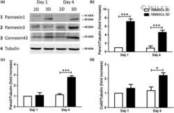
- 1 Immunolabeling for Panx1, Panx3, and Cx43 in HBMSCs in 2D or 3D cultures. (a-d) Panx1 immunolabeling at Day 1 and Day 4 in 2D and 3D cultures. (e-h) Panx3 immunolabeling at Days 1 and 4 in 2D and 3D cultures. (i-l) Cx43 immunolabeling at Days 1 and 4 in 2D and 3D cultures. In all cases, the primary antibody was detected with Alexa 488-conjugated IgG (in green) and nuclei stained by DAPI (blue)

- Submitted by
- Invitrogen Antibodies (provider)
- Main image

- Experimental details
- 6 Immunohistochemistry analysis of Pannexin1 and 3 in implanted matrices seeded with HBMSCs subcutaneously. Representative area of Panx1 (a-f) and Panx3 (g-l) immunohistochemistry of the transversal section of the tissue-engineering scaffolds after 3 and 8 weeks of implantation. Representative images of low magnification of the transversal sections (a,d,g,j) and higher magnifications of the periphery (Pe; b,e,h,k) and of the centre (Ce; c,f,i,l) of the implants. Representative histological images of Masson's trichrome (MT) staining (m,n,o) of subcutaneously implanted matrices loaded with HBMSCs, after 8 weeks of implantation, with a higher magnification at the periphery (n) and of the centre (o) of the implants
