 Explore
 Validate
 Learn
Learn Western blot
 Immunocytochemistry
Antibody data
- Antibody Data
- Antigen structure
- References [0]
- Comments [0]
- Validations
- Western blot [6]
- Chromatin Immunoprecipitation [1]
Submit
Validation data
Reference
Comment
Report error
- Product number
- PA5-29602 - Provider product page

- Provider
- Invitrogen Antibodies
- Product name
- Anti-Histone H3.3 Polyclonal Antibody
- Antibody type
- Polyclonal
- Antigen
- Recombinant protein fragment
- Description
- Recommended positive controls: Jurkat, Raji, K562, THP-1, HL-60, NCI-H929, NIH-3T3, JC, BCL-1, C2C12, Raw264.7, PC-12 , rice leaf. Predicted reactivity: Mouse (100%), Zebrafish (99%), Xenopus laevis (100%), Pig (100%), Rabbit (100%), Bovine (100%). Store product as a concentrated solution. Centrifuge briefly prior to opening the vial.
- Reactivity
- Human, Mouse, Rat
- Host
- Rabbit
- Isotype
- IgG
- Vial size
- 100 µL
- Concentration
- 1.74 mg/mL
- Storage
- Store at 4°C short term. For long term storage, store at -20°C, avoiding freeze/thaw cycles.
No comments: Submit comment
Supportive validation
- Submitted by
- Invitrogen Antibodies (provider)
- Main image

- Experimental details
- Western blot analysis of Histone H3.3 using 30 µg of A) Jurkat (B) Raji (C) K562 (D) THP-1 (E) HL-60 and F) NCI-H929 lysate. Samples were loaded onto a 15% SDS-PAGE gel and probed with a Histone H3.3 polyclonal antibody (Product # PA5-29602) at a dilution of 1:10,000.

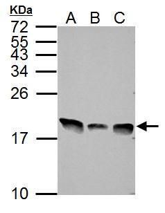
- Submitted by
- Invitrogen Antibodies (provider)
- Main image

- Experimental details
- Western blot analysis of Histone H3.3 using 30 µg of A) NIH-3T3 (B) JC and C) BCL-1 lysate. Samples were loaded onto a 15% SDS-PAGE gel and probed with a Histone H3.3 polyclonal antibody (Product # PA5-29602) at a dilution of 1:10,000.
- Submitted by
- Invitrogen Antibodies (provider)
- Main image

- Experimental details
- Western Blot analysis of Histone H3.3 was performed by separating 30 µg of various whole cell extracts by 15% SDS-PAGE. Proteins were transferred to a membrane and probed with a Histone H3.3 Polyclonal Antibody (Product # PA5-29602) at a dilution of 1:10000 and a HRP-conjugated anti-rabbit IgG secondary antibody.

- Submitted by
- Invitrogen Antibodies (provider)
- Main image
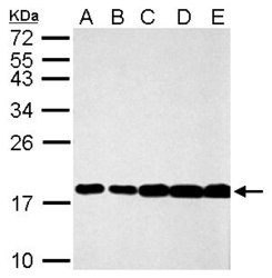
- Experimental details
- Western Blot analysis of Histone H3.3 was performed by separating 30 µg of various whole cell lysates by 15% SDS-PAGE. Proteins were transferred to a membrane and probed with a Histone H3.3 Polyclonal Antibody (Product # PA5-29602) at a dilution of 1:10000. The HRP-conjugated anti-rabbit IgG antibody was used to detect the primary antibody. A. NIH-3T3, B. JC, C. BCL-1, D. C2C12, E. Raw264.7.

- Submitted by
- Invitrogen Antibodies (provider)
- Main image

- Experimental details
- Western Blot analysis of Histone H3.3 was performed by separating 30 µg of PC-12 whole cell lysates by 15% SDS-PAGE. Proteins were transferred to a membrane and probed with a Histone H3.3 Polyclonal Antibody (Product # PA5-29602) at a dilution of 1:10000. The HRP-conjugated anti-rabbit IgG antibody was used to detect the primary antibody.

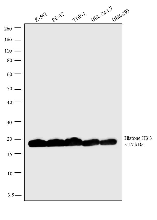
- Submitted by
- Invitrogen Antibodies (provider)
- Main image

- Experimental details
- Western blot analysis was performed on acid extracts (30µg lysate) of K-562 (Lane 1), PC-12 (Lane 2), THP-1 (Lane 3), HEL 92.1.7 (Lane 4), and HEK-293 (Lane 5). The blots were probed with Anti-Histone H3.3 Rabbit Polyclonal Antibody (Product # PA5-29602, 1:5000 dilution) and detected by chemiluminescence using Goat anti-Rabbit IgG (H+L) Superclonal™ Secondary Antibody, HRP conjugate (Product # A27036, 0.25µg/mL, 1:4000 dilution). A 17 kDa band corresponding to Histone H3.3 was observed across the cell lines tested. Known quantity of protein samples were electrophoresed using Novex® NuPAGE® 4-12 % Bis-Tris gel (Product # NP0321BOX), XCell SureLock™ Electrophoresis System (Product # EI0002) and Novex® Sharp Pre-Stained Protein Standard (Product # LC5800). Resolved proteins were then transferred onto a nitrocellulose membrane with iBlot® 2 Dry Blotting System (Product # IB21001). The membrane was probed with the relevant primary and secondary Antibody following blocking with 5 % skimmed milk. Chemiluminescent detection was performed using Pierce™ ECL Western Blotting Substrate (Product # 32106).
Supportive validation
- Submitted by
- Invitrogen Antibodies (provider)
- Main image

- Experimental details
- Enrichment of endogenous ANTI-HISTONE H3.3 protein at specific gene loci using Anti-ANTI-HISTONE H3.3 Rabbit Polyclonal Antibody: Chromatin Immunoprecipitation (ChIP) was performed using Anti-ANTI-HISTONE H3.3 Rabbit Polyclonal Antibody (Product # PA5-29602, 3 µg) on sheared chromatin from 2 million HeLa cells using the MAGnify ChIP system kit (Product # 49-2024). Normal Rabbit IgG was used as a negative IP control. The purified DNA was analyzed by 7500 Fast qPCR system (Product # 4351106) with optimized PCR primer pairs for the promoter of MYT-1 gene, SAT 2, Satellite alpha (SAT A) Repeat used as positive control target, and promoter of C-FOS, PABPC1 used as negative control target. Data is presented as fold enrichment of the antibody signal versus the negative control IgG using the comparative CT method.
