 Explore
 Validate
 Learn
Learn Western blot
 Immunohistochemistry
Antibody data
- Antibody Data
- Antigen structure
- References [1]
- Comments [0]
- Validations
- Western blot [11]
Submit
Validation data
Reference
Comment
Report error
- Product number
- PA5-17972 - Provider product page

- Provider
- Invitrogen Antibodies
- Product name
- MTM1 Polyclonal Antibody
- Antibody type
- Polyclonal
- Antigen
- Synthetic peptide
- Description
- This antibody is predicted to react with bovine, canine and mouse based on sequence homology.
- Reactivity
- Human, Mouse
- Host
- Goat
- Isotype
- IgG
- Vial size
- 100 µg
- Concentration
- 0.5 mg/mL
- Storage
- -20° C, Avoid Freeze/Thaw Cycles
Submitted references Pathogenic deep intronic MTM1 variant activates a pseudo-exon encoding a nonsense codon resulting in severe X-linked myotubular myopathy.
Bryen SJ, Oates EC, Evesson FJ, Lu JK, Waddell LB, Joshi H, Ryan MM, Cummings BB, McLean CA, MacArthur DG, Kornberg AJ, Cooper ST
European journal of human genetics : EJHG 2021 Jan;29(1):61-66
European journal of human genetics : EJHG 2021 Jan;29(1):61-66
No comments: Submit comment
Supportive validation
- Submitted by
- Invitrogen Antibodies (provider)
- Main image

- Experimental details
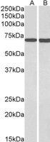
- Western Blot staining of HepG2 cell lysate using Product # PA5-17972 at a concentration of 0.3 µg/mL, the primary antibody incubation was 1 hour and the detection method was chemiluminescence.
- Submitted by
- Invitrogen Antibodies (provider)
- Main image

- Experimental details
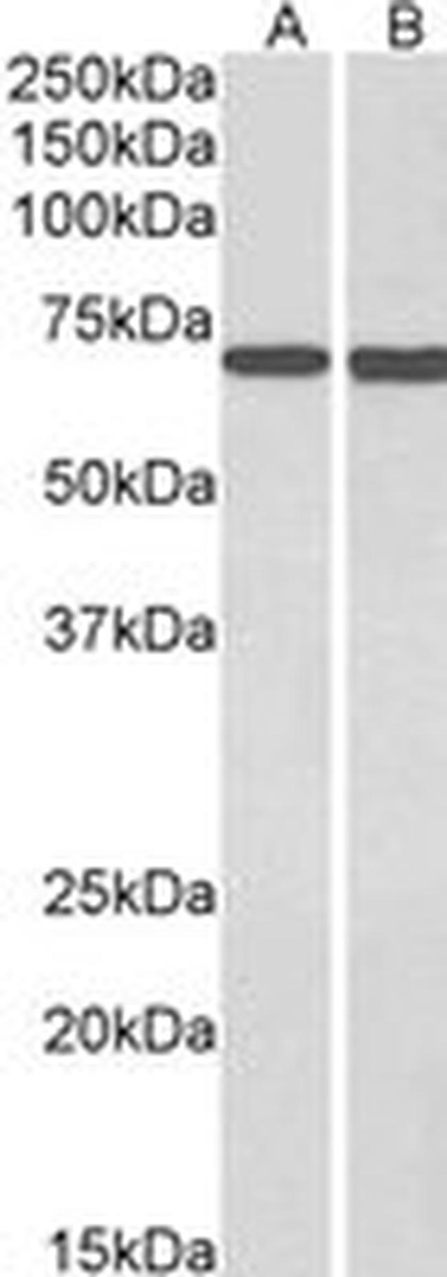
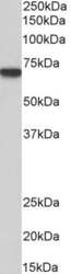
- Western blot analysis of MTM1 in Human Skeletal Muscle lysate (35µg protein in RIPA buffer). Samples were probed with the MTM1 antibody (Product # PA5-17972, 0.3µg/mL) for 1 hour. Western blot was detected by chemiluminescence.
- Submitted by
- Invitrogen Antibodies (provider)
- Main image

- Experimental details
- Western blot analysis of MTM1 in Human Skeletal Muscle lysate (35µg protein in RIPA buffer). Samples were probed with the MTM1 antibody (Product # PA5-17972, 0.3µg/mL) for 1 hour. Western blot was detected by chemiluminescence.
- Submitted by
- Invitrogen Antibodies (provider)
- Main image
- Experimental details
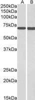
- Western blot analysis of MTM1 by a MTM1 monoclonal antibody (Product # PA5-17972) at a concentration of 1 µg/mL. HepG2 cell lysate (35µg protein in RIPA buffer). Primary incubation was 1 hour. Detected by chemiluminescence.

- Submitted by
- Invitrogen Antibodies (provider)
- Main image

- Experimental details
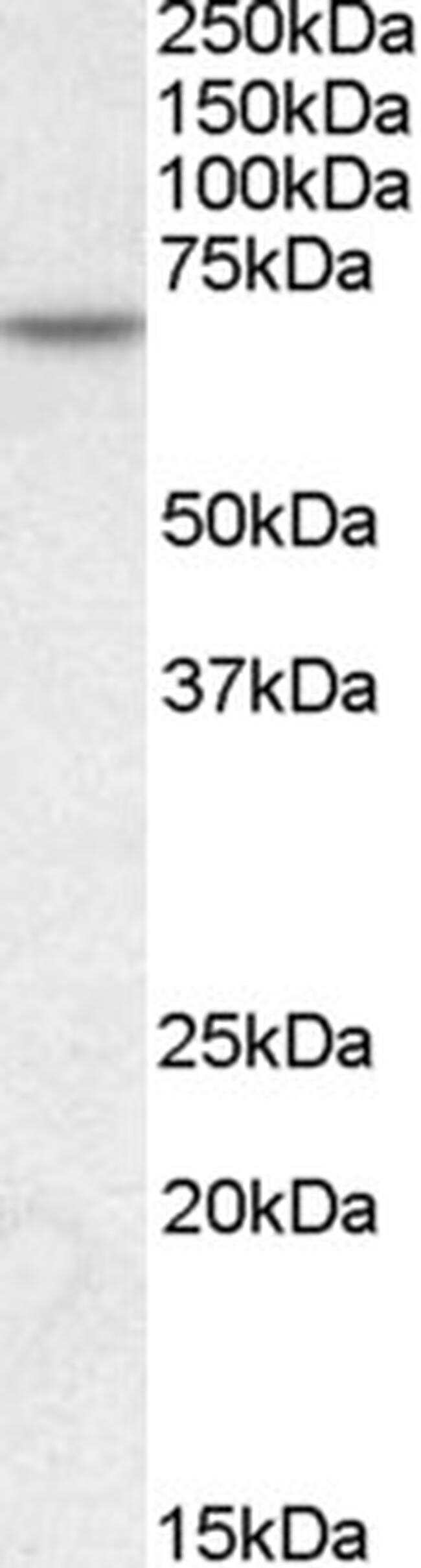
- Western blot analysis of MTM1 by a MTM1 monoclonal antibody (Product # PA5-17972) at a concentration of 1 µg/mL. Human Skeletal Muscle lysate (35µg protein in RIPA buffer). Primary incubation was 1 hour. Detected by chemiluminescence.
- Submitted by
- Invitrogen Antibodies (provider)
- Main image

- Experimental details
- Western Blot staining of HepG2 cell lysate using Product # PA5-17972 at a concentration of 0.3 µg/mL, the primary antibody incubation was 1 hour and the detection method was chemiluminescence.

- Submitted by
- Invitrogen Antibodies (provider)
- Main image

- Experimental details
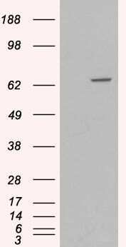
- Western blot of HEK293 overexpressing MTM1 using Product # PA5-17972, mock transfection as a control in first lane.
- Submitted by
- Invitrogen Antibodies (provider)
- Main image

- Experimental details
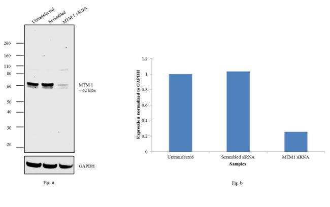
- Knockdown of MTM 1 was achieved by transfecting Hep G2 cells with MTM 1 specific siRNAs (Silencer® select Product # s9041). Western blot analysis (Fig. a) was performed using whole cell extracts from the MTM 1 knockdown cells (lane 3), non-specific scrambled siRNA transfected cells (lane 2) and untransfected cells (lane 1). The blots were probed with MTM 1 Polyclonal Antibody (Product # PA5-17972, 1:250 dilution) and Rabbit anti-Goat IgG (H+L) Superclonal™ Secondary Antibody, HRP conjugate (Product # A27014, 0.25 µg/mL, 1:4000 dilution). Densitometric analysis of this western blot is shown in histogram (Fig. b). Decrease in signal upon siRNA mediated knock down confirms that antibody is specific to MTM 1.
- Submitted by
- Invitrogen Antibodies (provider)
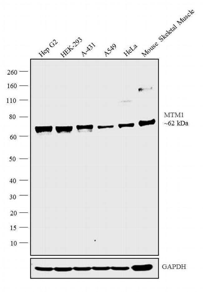
- Main image

- Experimental details
- Western blot analysis was performed membrane enriched extracts (30 µg lysate) of Hep G2 (Lane 1), HEK-293 (Lane 2), A-431 (Lane 3), A549 (Lane 4), HeLa (Lane 5) and tissue extract of Mouse Skeletal Muscle (Lane 6). The blot was probed with Anti-MTM1 antibody (Product # PA5-17972, 1µg/mL) and detected by chemiluminescence using Rabbit anti-Goat IgG (H+L) Superclonal™ Secondary Antibody, HRP conjugate (Product # A27014, 0.25 µg/mL, 1:4000 dilution). A 62 kDa band corresponding to MTM1 was observed across the cell lines and tissue tested.
- Submitted by
- Invitrogen Antibodies (provider)
- Main image

- Experimental details
- Western blot analysis of MTM1 by a MTM1 monoclonal antibody (Product # PA5-17972) at a concentration of 1 µg/mL. Mouse Skeletal Muscle lysate (35µg protein in RIPA buffer). Primary incubation was 1 hour. Detected by chemiluminescence.
