Explore
 Validate
 Learn
Learn Western blot
Antibody data
- Antibody Data
- Antigen structure
- References [1]
- Comments [0]
- Validations
- Western blot [3]
- Immunohistochemistry [3]
- Other assay [1]
Submit
Validation data
Reference
Comment
Report error
- Product number
- PA5-30763 - Provider product page

- Provider
- Invitrogen Antibodies
- Product name
- PPP2R2D Polyclonal Antibody
- Antibody type
- Polyclonal
- Antigen
- Recombinant protein fragment
- Description
- Recommended positive controls: IMR32, mouse brain, rat brain.
- Concentration
- 0.5 mg/mL
Submitted references CDC25B partners with PP2A to induce AMPK activation and tumor suppression in triple negative breast cancer.
Cairns J, Ly RC, Niu N, Kalari KR, Carlson EE, Wang L
NAR cancer 2020 Dec;2(4):zcaa039
NAR cancer 2020 Dec;2(4):zcaa039
No comments: Submit comment
Supportive validation
- Submitted by
- Invitrogen Antibodies (provider)
- Main image

- Experimental details
- Western blot analysis of PPP2R2D using 50 µg of mouse brain lysate. Samples were loaded onto a 10% SDS-PAGE gel and probed with a PPP2R2D polyclonal antibody (Product # PA5-30763) at a dilution of 1:500.

- Submitted by
- Invitrogen Antibodies (provider)
- Main image
- Experimental details
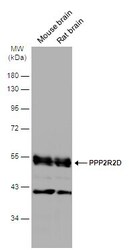
- Western blot analysis of PPP2R2D was performed by separating 50 µg of various tissue extracts by 10% SDS-PAGE. Proteins were transferred to a membrane and probed with a PPP2R2D Polyclonal Antibody (Product # PA5-30763) at a dilution of 1:5000. The HRP-conjugated anti-rabbit IgG antibody was used to detect the primary antibody.

- Submitted by
- Invitrogen Antibodies (provider)
- Main image
- Experimental details
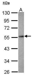
- Western Blot using PPP2R2D Polyclonal Antibody (Product # PA5-30763). Sample (30 µg of whole cell lysate). Lane A:IMR32. 10% SDS PAGE. PPP2R2D Polyclonal Antibody (Product # PA5-30763) diluted at 1:1,000. The HRP-conjugated anti-rabbit IgG antibody was used to detect the primary antibody.

Supportive validation
- Submitted by
- Invitrogen Antibodies (provider)
- Main image

- Experimental details
- Immunohistochemistry (Paraffin) analysis of PPP2R2D was performed in paraffin-embedded mouse testis tissue using PPP2R2D Polyclonal Antibody (Product # PA5-30763) at a dilution of 1:500.

- Submitted by
- Invitrogen Antibodies (provider)
- Main image

- Experimental details
- Immunohistochemistry (Paraffin) analysis of PPP2R2D was performed in paraffin-embedded mouse duodenum tissue using PPP2R2D Polyclonal Antibody (Product # PA5-30763) at a dilution of 1:500.

- Submitted by
- Invitrogen Antibodies (provider)
- Main image

- Experimental details
- Immunohistochemistry (Paraffin) analysis of PPP2R2D was performed in paraffin-embedded rat testis tissue using PPP2R2D Polyclonal Antibody (Product # PA5-30763) at a dilution of 1:500.

Supportive validation
- Submitted by
- Invitrogen Antibodies (provider)
- Main image

- Experimental details
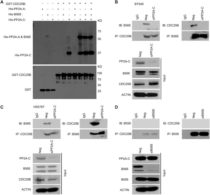
- Figure 3. CDC25B interacts directly with the PP2A-C subunit. ( A ) Top: GST-CDC25B was used to pull down purified His-PP2A-A, His-B56delta, His-PP2A-C, His-PP2A-A + B56delta, His-PP2A-A + PP2A-C, His-B56delta + PP2A-C or His-PP2A-A + B56delta + PP2A-C. Bottom: GST immunoblot of SDS-PAGE gel demonstrating the amount of CDC25B used for pull-down experiments. ( B and C ) The CDC25B-B56delta interaction in Neg or PP2A-C KD cells. BT549 and HS578T cells were transfected with Neg or siPP2A-C. Forty-eight hours later, cell lysates were subjected to IP with control IgG or anti-CDC25B antibody examining the CDC25B-B56delta interaction. Reciprocal IP with B56delta antibody was also performed to examine the CDC25B-B56delta interaction. The immunoprecipitates were blotted with the indicated antibodies. ( D ) B56delta, not B55delta, interacts with CDC25B. BT549 cells were transfected with siB56delta. Forty-eight hours later, cell lysates were subjected to IP with control IgG or anti-CDC25B antibody examining the CDC25B-B55delta interaction. Reciprocal IP with B55delta antibody was also performed to exam the CDC25B-B55delta interaction. The immunoprecipitates were blotted with the indicated antibodies. Western blot analysis demonstrates the protein level of B55delta in Neg or B56delta KD cells. Data information: all data presented are a representation of N = 3 independent experiments.