 Explore
 Validate
 Learn
Learn ELISA
ELISAAntibody data
- Antibody Data
- Antigen structure
- References [3]
- Comments [0]
- Validations
- ELISA [1]
- Flow cytometry [2]
Submit
Validation data
Reference
Comment
Report error
- Product number
- MAB7144 - Provider product page

- Provider
- R&D Systems
- Product name
- Human B7-H6 Antibody
- Antibody type
- Monoclonal
- Description
- Protein A or G purified from hybridoma culture supernatant. Detects human B7-H6 in ELISA.
- Reactivity
- Human
- Host
- Mouse
- Conjugate
- Unconjugated
- Antigen sequence
Q68D85- Isotype
- IgG
- Antibody clone number
- 875001
- Vial size
- 100 ug
- Concentration
- LYOPH
- Storage
- Use a manual defrost freezer and avoid repeated freeze-thaw cycles. 12 months from date of receipt, -20 to -70 °C as supplied. 1 month, 2 to 8 °C under sterile conditions after reconstitution. 6 months, -20 to -70 °C under sterile conditions after reconstitution.
Submitted references B7H6 is a functional ligand for NKp30 in rat and cattle and determines NKp30 reactivity toward human cancer cell lines.
Control of immune ligands by members of a cytomegalovirus gene expansion suppresses natural killer cell activation.
Human cytomegalovirus escapes immune recognition by NK cells through the downregulation of B7-H6 by the viral genes US18 and US20.
Bjørnsen EG, Thiruchelvam-Kyle L, Hoelsbrekken SE, Henden C, Saether PC, Boysen P, Daws MR, Dissen E
European journal of immunology 2019 Jan;49(1):54-65
European journal of immunology 2019 Jan;49(1):54-65
Control of immune ligands by members of a cytomegalovirus gene expansion suppresses natural killer cell activation.
Fielding CA, Weekes MP, Nobre LV, Ruckova E, Wilkie GS, Paulo JA, Chang C, Suárez NM, Davies JA, Antrobus R, Stanton RJ, Aicheler RJ, Nichols H, Vojtesek B, Trowsdale J, Davison AJ, Gygi SP, Tomasec P, Lehner PJ, Wilkinson GW
eLife 2017 Feb 10;6
eLife 2017 Feb 10;6
Human cytomegalovirus escapes immune recognition by NK cells through the downregulation of B7-H6 by the viral genes US18 and US20.
Charpak-Amikam Y, Kubsch T, Seidel E, Oiknine-Djian E, Cavaletto N, Yamin R, Schmiedel D, Wolf D, Gribaudo G, Messerle M, Cicin-Sain L, Mandelboim O
Scientific reports 2017 Aug 17;7(1):8661
Scientific reports 2017 Aug 17;7(1):8661
No comments: Submit comment
Supportive validation
- Submitted by
- R&D Systems (provider)
- Main image
- Experimental details
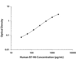
- Human B7-H6 ELISA Standard Curve. Recombinant Human B7-H6 protein was serially diluted 2-fold and captured by Mouse Anti-Human B7-H6 Monoclonal Antibody (Catalog # MAB7144) coated on a Clear Polystyrene Microplate (Catalog # DY990). Mouse Anti-Human B7-H6 Monoclonal Antibody (Catalog # MAB71442) was biotinylated and incubated with the protein captured on the plate. Detection of the standard curve was achieved by incubating Streptavidin-HRP (Catalog # DY998) followed by Substrate Solution (Catalog # DY999) and stopping the enzymatic reaction with Stop Solution (Catalog # DY994).

Supportive validation
- Submitted by
- R&D Systems (provider)
- Main image

- Experimental details
- Detection of B7-H6 in HeLa Human Cell Line by Flow Cytometry. HeLa human cervical epithelial carcinoma cell line was stained with Mouse Anti-Human B7-H6 Monoclonal Antibody (Catalog # MAB7144, filled histogram) or isotype control antibody (Catalog # MAB002, open histogram), followed by Allophycocyanin-conjugated Anti-Mouse IgG Secondary Antibody (Catalog # F0101B).

- Submitted by
- R&D Systems (provider)
- Main image

- Experimental details
- B7-H6 Specificity is Shown by Flow Cytometry in Knockout Cell Line. B7-H6 knockout HeLa human cervical epithelial carcinoma cell line was stained with Mouse Anti-Human B7-H6 Monoclonal Antibody (Catalog # MAB7144, filled histogram) or isotype control antibody (Catalog # MAB002, open histogram) followed by anti-Mouse IgG PE-conjugated secondary antibody (Catalog # F0102B). No staining in the B7-H6 knockout HeLa cell line was observed. View our protocol for Staining Membrane-associated Proteins.
