 Explore
 Validate
 Learn
Learn Western blot
 Immunocytochemistry
Antibody data
- Antibody Data
- Antigen structure
- References [1]
- Comments [0]
- Validations
- Immunocytochemistry [1]
- Immunohistochemistry [5]
Submit
Validation data
Reference
Comment
Report error
- Product number
- HPA000898 - Provider product page

- Provider
- Atlas Antibodies
- Proper citation
- Atlas Antibodies Cat#HPA000898, RRID:AB_1079032
- Product name
- Anti-HSPA9
- Antibody type
- Polyclonal
- Reactivity
- Human, Mouse, Rat
- Host
- Rabbit
- Conjugate
- Unconjugated
- Antigen sequence
GIVHVSAKDKGTGREQQIVIQSSGGLSKDDIENMV
KNAEKYAEEDRRKKERVEAVNMAEGIIHDTETKME
EFKDQLPADECNKLKEEISKMRELLARKDSETGEN
IRQAASSLQQASLKLFEMAYKKMASEREGSGSSGT- Isotype
- IgG
- Vial size
- 100 µl
- Storage
- Store at +4°C for short term storage. Long time storage is recommended at -20°C.
Submitted references Comparative proteomics analysis of gastric cancer stem cells.
Morisaki T, Yashiro M, Kakehashi A, Inagaki A, Kinoshita H, Fukuoka T, Kasashima H, Masuda G, Sakurai K, Kubo N, Muguruma K, Ohira M, Wanibuchi H, Hirakawa K
PloS one 2014;9(11):e110736
PloS one 2014;9(11):e110736
No comments: Submit comment
Supportive validation
- Submitted by
- Atlas Antibodies (provider)
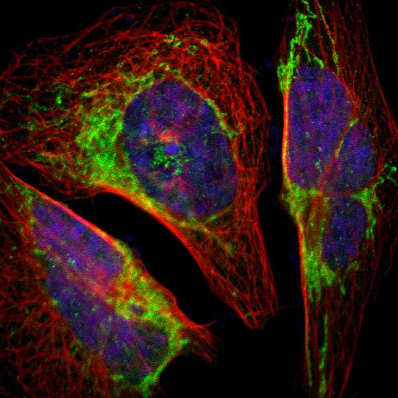
- Main image

- Experimental details
- Immunofluorescent staining of human cell line U-2 OS shows localization to mitochondria.
- Sample type
- HUMAN
Supportive validation
- Submitted by
- Atlas Antibodies (provider)
- Main image
- Experimental details
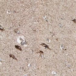
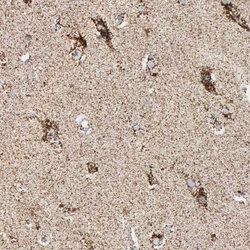
- Immunohistochemical staining of human cerebral cortex shows strong cytoplasmic positivity in neuronal cells.
- Submitted by
- Atlas Antibodies (provider)
- Main image
- Experimental details
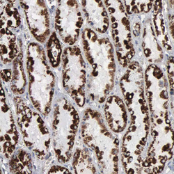
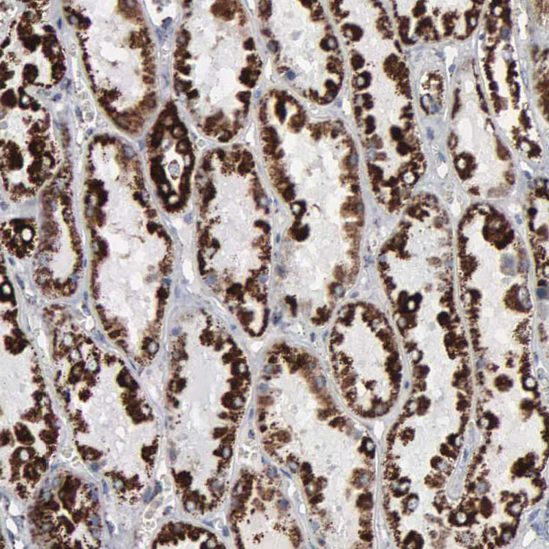
- Immunohistochemical staining of human kidney shows strong positivity in mitochondria in cells in tubules.
- Sample type
- HUMAN
- Submitted by
- Atlas Antibodies (provider)
- Main image
- Experimental details
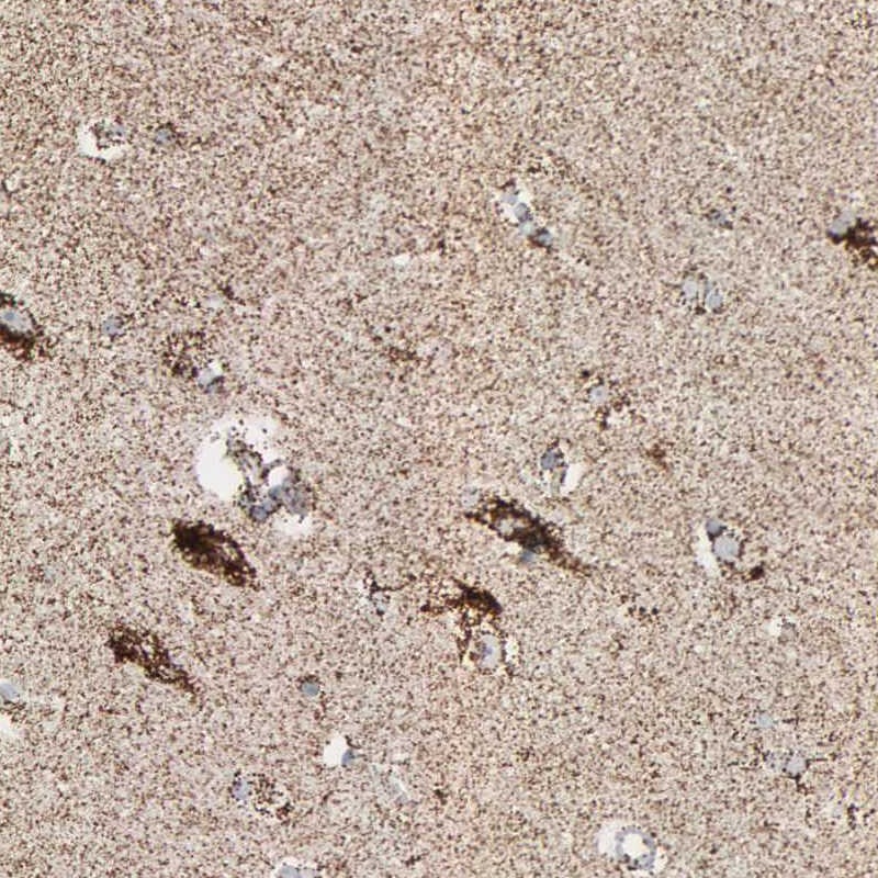
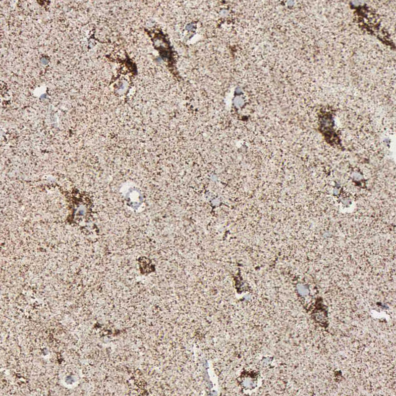
- Immunohistochemical staining of human cerebral cortex shows strong positivity in mitochondria in neurons.
- Sample type
- HUMAN
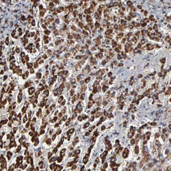
- Submitted by
- Atlas Antibodies (provider)
- Main image
- Experimental details
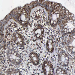
- Immunohistochemical staining of human colon shows strong positivity in mitochondria in glandular cells.
- Sample type
- HUMAN

- Submitted by
- Atlas Antibodies (provider)
- Main image
- Experimental details
- Immunohistochemical staining of human liver shows strong positivity in mitochondria in hepatocytes.
- Sample type
- HUMAN
