 Explore
 Validate
 Learn
Learn Western blot
Antibody data
- Antibody Data
- Antigen structure
- References [1]
- Comments [0]
- Validations
- Western blot [6]
- Immunohistochemistry [1]
- Other assay [1]
Submit
Validation data
Reference
Comment
Report error
- Product number
- PA5-21495 - Provider product page

- Provider
- Invitrogen Antibodies
- Product name
- VAV1 Polyclonal Antibody
- Antibody type
- Polyclonal
- Antigen
- Recombinant protein fragment
- Description
- Recommended positive controls: Jurkat, NCI-H929, BCL-1.
- Concentration
- 0.49 mg/mL
Submitted references Expression of P-REX2a is associated with poor prognosis in endometrial malignancies.
Takeshita S, Yamashita Y, Shiomi K, Suzuki N, Yoshida J, Naiki-Ito A, Suzuki S, Akatsuka S, Toyokuni S, Takahashi T, Mase S, Arakawa A, Sugiura-Ogasawara M, Takahashi S
Oncotarget 2018 May 15;9(37):24778-24786
Oncotarget 2018 May 15;9(37):24778-24786
No comments: Submit comment
Supportive validation
- Submitted by
- Invitrogen Antibodies (provider)
- Main image

- Experimental details
- Western blot analysis of VAV1 using 30 µg of A) MOLT4 and B) Raji lysate. Samples were loaded onto a 7.5% SDS-PAGE gel and probed with a VAV1 polyclonal antibody (Product # PA5-21495) at a dilution of 1:500.

- Submitted by
- Invitrogen Antibodies (provider)
- Main image

- Experimental details
- Western Blot using VAV1 Polyclonal Antibody (Product # PA5-21495). Various whole cell extracts (30 µg) were separated by 7.5% SDS-PAGE, and the membrane was blotted with VAV1 Polyclonal Antibody (Product # PA5-21495) diluted at 1:500. The HRP-conjugated anti-rabbit IgG antibody was used to detect the primary antibody.

- Submitted by
- Invitrogen Antibodies (provider)
- Main image

- Experimental details
- Western Blot analysis of VAV1 was performed by separating 30 µg of various whole cell extracts by 7.5% SDS-PAGE. Proteins were transferred to a membrane and probed with a VAV1 Polyclonal Antibody (Product # PA5-21495) at a dilution of 1:500 and a HRP-conjugated anti-rabbit IgG secondary antibody.

- Submitted by
- Invitrogen Antibodies (provider)
- Main image

- Experimental details
- Western Blot using VAV1 Polyclonal Antibody (Product # PA5-21495). Various whole cell extracts (30 µg) were separated by 7.5% SDS-PAGE, and the membrane was blotted with VAV1 Polyclonal Antibody (Product # PA5-21495) diluted at 1:500. The HRP-conjugated anti-rabbit IgG antibody was used to detect the primary antibody.

- Submitted by
- Invitrogen Antibodies (provider)
- Main image

- Experimental details
- VAV1 Polyclonal Antibody detects VAV1 protein by western blot analysis. A. 30 µg BCL-1 whole cell lysate/extract.7.5% SDS-PAGE. VAV1 Polyclonal Antibody (Product # PA5-21495) dilution: 1:1,000. The HRP-conjugated anti-rabbit IgG antibody was used to detect the primary antibody.

- Submitted by
- Invitrogen Antibodies (provider)
- Main image

- Experimental details
- Western blot was performed using Anti-VAV1 Polyclonal Antibody (Product # PA5-21495) and a 100kDa band corresponding to VAV1 was observed in Raji, MOLT-4, Jurkat, THP-1, Mouse Thymus, and not in Mouse Kidney which is reported negative for VAV1 expression. Whole cell lysates (30 µg lysate) of Raji (Lane 1), MOLT-4 (Lane 2), Jurkat (Lane 3), THP-1 (Lane 4), tissue extracts (30 µg lysate) of Mouse Thymus (Lane 5) and Mouse Kidney (Lane 6) were electrophoresed using NuPAGE® 10 % Bis-Tris gel (Product # NP0302BOX). Resolved proteins were then transferred onto a nitrocellulose membrane (Product # IB23001) by iBlot® 2 Dry Blotting System (Product # IB21001). The blot was probed with the primary antibody (1:1000 dilution) and detected by chemiluminescence Goat Anti-Rabbit IgG Secondary Antibody, HRP conjugate (Product # A27036, 1:4000 dilution) using the iBright FL 1000 (Product # A32752). Chemiluminescent detection was performed using SuperSignal™ West Dura Extended Duration Substrate (Product # 34076)..

Supportive validation
- Submitted by
- Invitrogen Antibodies (provider)
- Main image
- Experimental details
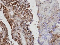
- Immunohistochemical analysis of paraffin-embedded human serous ovarian cancer, using VAV1 (Product # PA5-21495) antibody at 1:100 dilution. Antigen Retrieval: EDTA based buffer, pH 8.0, 15 min.

Supportive validation
- Submitted by
- Invitrogen Antibodies (provider)
- Main image

- Experimental details
- Figure 3 ( A ) Pathway analysis of 1,882 genes that displayed elevated expression levels by more than 2-fold and p -values less than 0.05 compared to the P-REX2a-knockdown groups and control using GeneSpring GX 13.1 software. Pathways with matched entities with p -values less than 0.05 are shown. ( B ) Vav1 knockdown experiments using OMC-2 and JHUEM-14 cells. Quantitative reverse-transcription PCR shows effective knockdown of Vav1 in both cell lines with 2 different siRNA sequences (left), but no significant changes are observed in migration assays (right). RT-PCR, reverse-transcription PCR; ** P < 0.01, * P < 0.05, n.s, not significant; p >= 0.05. ( C ) Immunohistochemistry for Vav 1. A case of negative immunostaining is shown on the upper left. Positive cytoplasmic immunostaining was further quantified and scored into 3 levels, 1+ to 3+. See Materials & Methods for the criteria of the immunostaining. Scale bar (black line); 100 mum.
