Explore
 Validate
 Learn
Learn Western blot
Antibody data
- Antibody Data
- Antigen structure
- References [7]
- Comments [0]
- Validations
- Western blot [2]
- Other assay [7]
Submit
Validation data
Reference
Comment
Report error
- Product number
- 40-2000 - Provider product page

- Provider
- Invitrogen Antibodies
- Product name
- HOXB7 Polyclonal Antibody
- Antibody type
- Polyclonal
- Antigen
- Synthetic peptide
- Reactivity
- Human
- Host
- Rabbit
- Isotype
- IgG
- Vial size
- 100 µg
- Concentration
- 0.25 mg/mL
- Storage
- -20°C
Submitted references Silencing of hsa_circ_0009035 Suppresses Cervical Cancer Progression and Enhances Radiosensitivity through MicroRNA 889-3p-Dependent Regulation of HOXB7.
Long non-coding RNA SNHG8 promotes prostate cancer progression through repressing miR-384 and up-regulating HOXB7.
Psoralen Suppresses Cisplatin-Mediated Resistance and Induces Apoptosis of Gastric Adenocarcinoma by Disruption of the miR196a-HOXB7-HER2 Axis.
Targeting HOX/PBX dimer formation as a potential therapeutic option in esophageal squamous cell carcinoma.
The roles of HOXB7 in promoting migration, invasion, and anti-apoptosis in gastric cancer.
HOXB7-S3 inhibits the proliferation and invasion of MCF-7 human breast cancer cells.
HOXB7 expression by myeloma cells regulates their pro-angiogenic properties in multiple myeloma patients.
Zhao X, Dong W, Luo G, Xie J, Liu J, Yu F
Molecular and cellular biology 2021 May 21;41(6):e0063120
Molecular and cellular biology 2021 May 21;41(6):e0063120
Long non-coding RNA SNHG8 promotes prostate cancer progression through repressing miR-384 and up-regulating HOXB7.
Shi Z, Zhang H, Jie S, Yang X, Huang Q, Mao Y, Zhang Y
The journal of gene medicine 2021 Mar;23(3):e3309
The journal of gene medicine 2021 Mar;23(3):e3309
Psoralen Suppresses Cisplatin-Mediated Resistance and Induces Apoptosis of Gastric Adenocarcinoma by Disruption of the miR196a-HOXB7-HER2 Axis.
Jin L, Ma XM, Wang TT, Yang Y, Zhang N, Zeng N, Bai ZG, Yin J, Zhang J, Ding GQ, Zhang ZT
Cancer management and research 2020;12:2803-2827
Cancer management and research 2020;12:2803-2827
Targeting HOX/PBX dimer formation as a potential therapeutic option in esophageal squamous cell carcinoma.
Shen LY, Zhou T, Du YB, Shi Q, Chen KN
Cancer science 2019 May;110(5):1735-1745
Cancer science 2019 May;110(5):1735-1745
The roles of HOXB7 in promoting migration, invasion, and anti-apoptosis in gastric cancer.
Joo MK, Park JJ, Yoo HS, Lee BJ, Chun HJ, Lee SW, Bak YT
Journal of gastroenterology and hepatology 2016 Oct;31(10):1717-1726
Journal of gastroenterology and hepatology 2016 Oct;31(10):1717-1726
HOXB7-S3 inhibits the proliferation and invasion of MCF-7 human breast cancer cells.
Ma R, Zhang D, Hu PC, Li Q, Lin CY
Molecular medicine reports 2015 Oct;12(4):4901-8
Molecular medicine reports 2015 Oct;12(4):4901-8
HOXB7 expression by myeloma cells regulates their pro-angiogenic properties in multiple myeloma patients.
Storti P, Donofrio G, Colla S, Airoldi I, Bolzoni M, Agnelli L, Abeltino M, Todoerti K, Lazzaretti M, Mancini C, Ribatti D, Bonomini S, Franceschi V, Pistoia V, Lisignoli G, Pedrazzini A, Cavicchi O, Neri A, Rizzoli V, Giuliani N
Leukemia 2011 Mar;25(3):527-37
Leukemia 2011 Mar;25(3):527-37
No comments: Submit comment
Supportive validation
- Submitted by
- Invitrogen Antibodies (provider)
- Main image

- Experimental details
- Western Blot analysis of (A) A375, (B) NTERA (NT2/D1), (C) SK-MEL-19, (D) SK-MEL-37, (E) KARPAS 299, (F) SW480, and (G) HT-29 cell lysates using Zymed Rb anti-HOXB7 (Product # 40-2000).

- Submitted by
- Invitrogen Antibodies (provider)
- Main image

- Experimental details
- Western blot analysis of SW480 cell lysates using Rb anti-HOXB7 (Product # 40-2000).

Supportive validation
- Submitted by
- Invitrogen Antibodies (provider)
- Main image

- Experimental details
- 5 FIGURE SNHG8 up-regulates HOXB7 expression via adsorbing miR-384. (A) The binding site between HOXB7 3'-UTR and miR-384 was predicted by the StarBase database. (B) A dual-luciferase reporter experiment was conducted to detect the binding relationship between HOXB7 and miR-384. (C) Western blotting was performed to detect HOXB7 protein expression in the PCa cell line and human prostate epithelial cells. (D) Western blotting was performed to detect the effect of si-SNHG8 # 1 and miR-384 inhibitors on HOXB7 protein expression. (E-G) qRT-PCR was carried out to detect HOXB7 mRNA expression in PCa tissues (E) and the correlations between HOXB7 and miR-384 (F) and HOXB7 and SNHG8 expression (G) were analyzed. *** p < 0.001

- Submitted by
- Invitrogen Antibodies (provider)
- Main image

- Experimental details
- Figure 5 The target gene of miR-196a-5p is HOXB7. ( A ) The predicted results of miR-196a-5p and HOXB7 in TargetScan. ( B ) In dual-luciferase reporter assays, miR-196a-5p suppressed the luciferase activity of the HOXB7-WT vector, and the luciferase activity in the MUT-group (HOXB7-MUT + miR-196a-5p mimic) was greatly higher than that in the WT-group (HOXB7-WT + miR-196a-5p mimic) in MGC803 cell line. ( C ) The relative HOXB7 expression between MGC803 cell line and MGC803/DDP cell line was analyzed by RT-qPCR. ( D and E ) The relative expression of HOXB7 between MGC803 cell line and MGC803/DDP cell line was measured by WB. ( F ) The relative expression of HOXB7 following transfection with the three different concentrations of miR-196a-5p inhibitor, compared to the miR-196a-5p inhibitor control, in MGC803 cell line was analyzed by RT-qPCR. ( G and H ) The relative expression of HOXB7 in MGC803 cells following transfection with the miR-196a-5p inhibitor and the miR-196a-5p inhibitor control was measured by WB. ( I ) The relative expression of HOXB7 following transfection with the three different concentrations of miR-196a-5p mimic, compared with the miR-196a-5p mimic control, in MGC803/DDP cell line was analyzed by RT-qPCR. ( J and K ) WB results of HOXB7 expression levels in MGC803/DDP cell line after transfected with the miR-196a-5p mimic and the miR-196a-5p mimic control. ( L ) Kaplan-Meier survival curves demonstrated lower HOXB7 levels (n=394) were relevant with higher pat

- Submitted by
- Invitrogen Antibodies (provider)
- Main image

- Experimental details
- Figure 10 Psoralen could reverse cisplatin resistance of GC cells via activating the miR196a-HOXB7-HER2 Pathway. ( A ) Western blot results of HOXB7, HER2, Bcl-2 and CyclinD1 protein levels between MGC803 cells and MGC803/DDP cells following cultured with psoralen and the negative control. ( B-E ) The relative expression of HOXB7 ( B ), HER2 ( C ), Bcl-2 ( D ) and CyclinD1 ( E ) protein between MGC803 cells and MGC803/DDP cells following cultured with psoralen and the negative control was measured by WB. The beta-actin antibody was utilized for internal reference. Each assay was conducted in triplicate. ****P < 0.0001, ***P < 0.001 and mean+-SD were utilized to show the data.

- Submitted by
- Invitrogen Antibodies (provider)
- Main image

- Experimental details
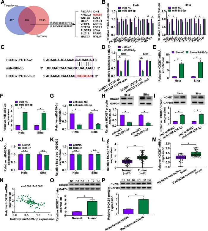
- FIG 7 HOXB7 was a direct target of miR-889-3p in CC cells. (A) Venn diagram showing the putative targets of miR-889-3p predicted by Targetscan and Starbase softwares. (B) qRT-PCR analysis of genes in HeLa cells transfected with the miR-NC mimic or miR-889-3p mimic. (C) Schematic of the target sequence for miR-889-3p identified by Starbase software and the mutant of the seed region. (D) Dual-luciferase reporter assays in both HeLa and Siha cells. (E) RNA pulldown assays in HeLa and Siha cells using Bio-NC or Bio-miR-889-3p. Relative miR-889-3p expression by qRT-PCR in cells transfected with an miR-NC mimic, an miR-889-3p mimic (F), anti-miR-NC, or anti-miR-889-3p (G). HOXB7 protein level determined by Western blotting in cells transfected with an miR-NC mimic, an miR-889-3p mimic (H), anti-miR-NC, or anti-miR-889-3p (I). (J and K) qRT-PCR analysis of miR-889-3p and hsa_circ_0009035 expression levels in cells transfected with a negative-control plasmid (pcDNA) or a HOXB7-overexpressing plasmid (HOXB7). Relative HOXB7 expression determined by qRT-PCR in 82 pairs of CC tissues and adjacent normal tissues (L), CC tissues from 36 primary patients (defined as radiation-sensitive CC) and 46 recurrent patients after radiation treatment (defined as radiation-resistant CC) (M). (N) Correlation between HOXB7 mRNA and miR-889-3p expression levels in CC tissues using the Spearman test. HOXB7 protein expression in 3 pairs of CC tissues and adjacent normal tissues (O), CC tissues from 3 prim
- Submitted by
- Invitrogen Antibodies (provider)
- Main image
- Experimental details
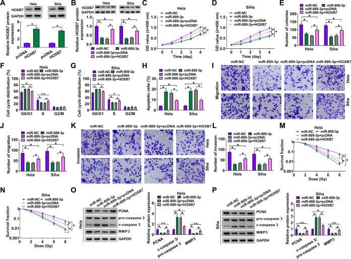
- FIG 8 The effects of miR-889-3p overexpression on CC progression and radiosensitivity in vitro were mediated by HOXB7. (A) Relative HOXB7 protein level determined by Western blotting in cells transfected with a negative-control plasmid (pcDNA) or a HOXB7-overexpressing plasmid (HOXB7). HeLa and Siha cells were transfected with an miR-NC mimic, an miR-899-3p mimic, an miR-899-3p mimic plus pcDNA, or an miR-899-3p mimic plus HOXB7, followed by the determination of HOXB7 protein level by Western blotting (B), cell proliferation by CCK-8 assay (C and D), analysis of cell colony formation by colony formation assay (E), analysis of cell cycle progression and apoptosis by flow cytometry (F to H), analysis of cell migration and invasion by transwell assay (I to L), determination of cell survival fraction by colony formation upon radiation exposure (M and N), and measurement of the levels of PCNA, c-caspase 3, procaspase 3, and MMP3 by Western blotting (O and P). * , P < 0.05.
- Submitted by
- Invitrogen Antibodies (provider)
- Main image
- Experimental details
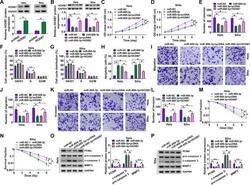
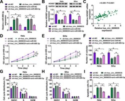
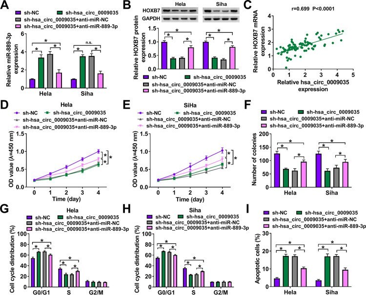
- FIG 9 hsa_circ_0009035 modulated HOXB7 expression and CC progression in vitro by miR-889-3p. Expression levels of miR-889-3p (A) and HOXB7 protein (B) in sh-NC-infected or sh-hsa_circ_0009035-transduced cells transfected with or without anti-miR-NC or anti-miR-889-3p. (C) Correlation between HOXB7 mRNA and hsa_circ_0009035 expression levels in CC tissues using the Spearman test. sh-NC-infected or sh-hsa_circ_0009035-transduced HeLa and Siha cells were transfected with or without anti-miR-NC or anti-miR-889-3p, followed by the assessment of cell proliferation by CCK-8 assay (D and E), cell colony formation by colony formation assay (F), and cell cycle progression and apoptosis by flow cytometry (G to I). * , P < 0.05.
- Submitted by
- Invitrogen Antibodies (provider)
- Main image
- Experimental details
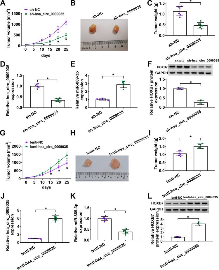
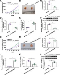
- FIG 11 hsa_circ_0009035 regulated tumor growth in vivo . (A) Growth curves of the xenograft tumors formed by sh-NC-infected or sh-hsa_circ_0009035-transduced HeLa cells ( n = 6 per group). Representative images (B), tumor average weight (C), hsa_circ_0009035 (D) and miR-889-3p (E) levels determined by qRT-PCR, and HOXB7 protein expression determined by Western blotting (F) of the xenograft tumors formed by HeLa cells infected with sh-NC or sh-hsa_circ_0009035, on day 25 after subcutaneous injection ( n = 6 per group). (G) Growth curves of the xenograft tumors formed by lenti-NC-infected or lenti-hsa_circ_0009035-transduced HeLa cells ( n = 6 per group). Representative images (H), tumor average weight (I), hsa_circ_0009035 (J), and miR-889-3p (K) levels determined by qRT-PCR, and HOXB7 protein expression determined by Western blotting (L) of the xenograft tumors formed by HeLa cells infected with lenti-NC or lenti-hsa_circ_0009035, on day 25 after subcutaneous injection ( n = 6 per group). * , P < 0.05.