 Explore
 Validate
 Learn
Learn Western blot
Antibody data
- Antibody Data
- Antigen structure
- References [1]
- Comments [0]
- Validations
- Western blot [1]
- Immunocytochemistry [1]
Submit
Validation data
Reference
Comment
Report error
- Product number
- AF3919 - Provider product page

- Provider
- R&D Systems
- Product name
- Human IRAK4 Antibody
- Antibody type
- Polyclonal
- Description
- Antigen Affinity-purified. Detects human IRAK4 in Western blots. In Western blots, less than 1% cross-reactivity with recombinant human (rh) IRAK1 and rhIRAK2 is observed.
- Reactivity
- Human
- Host
- Goat
- Conjugate
- Unconjugated
- Antigen sequence
Q9NWZ3- Isotype
- IgG
- Vial size
- 100 ug
- Concentration
- LYOPH
- Storage
- Use a manual defrost freezer and avoid repeated freeze-thaw cycles. 12 months from date of receipt, -20 to -70 °C as supplied. 1 month, 2 to 8 °C under sterile conditions after reconstitution. 6 months, -20 to -70 °C under sterile conditions after reconstitution.
Submitted references Contribution of globular death domains and unstructured linkers to MyD88.IRAK-4 heterodimer formation: an explanation for the antagonistic activity of MyD88s.
Mendoza-Barberá E, Corral-Rodríguez MA, Soares-Schanoski A, Velarde M, Macieira S, Messerschmidt A, López-Collazo E, Fuentes-Prior P
Biochemical and biophysical research communications 2009 Feb 27;380(1):183-7
Biochemical and biophysical research communications 2009 Feb 27;380(1):183-7
No comments: Submit comment
Supportive validation
- Submitted by
- R&D Systems (provider)
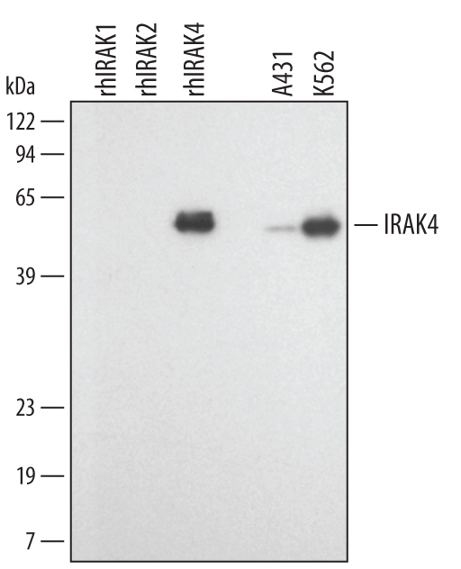
- Main image

- Experimental details
- Detection of Human IRAK4 by Western Blot. Western blot shows lysates of A431 human epithelial carcinoma cell line and K562 human chronic myelogenous leukemia cell line. PVDF membrane was probed with 1 µg/mL Goat Anti-Human IRAK4 Antigen Affinity-purified Polyclonal Antibody (Catalog # AF3919) followed by HRP-conjugated Anti-Goat IgG Secondary Antibody (Catalog # HAF109). For additional reference, recombinant human IRAK1, IRAK2, and IRAK4 (2 ng/lane) were included. A specific band for IRAK4 was detected at approximately 55 kDa (as indicated). This experiment was conducted under reducing conditions and using Immunoblot Buffer Group 1.
Supportive validation
- Submitted by
- R&D Systems (provider)
- Main image

- Experimental details
- IRAK4 in THP-1 Human Cell Line. IRAK4 was detected in immersion fixed THP-1 human acute monocytic leukemia cell line using Goat Anti-Human IRAK4 Antigen Affinity-purified Polyclonal Antibody (Catalog # AF3919) at 15 µg/mL for 3 hours at room temperature. Cells were stained using the NorthernLights™ 557-conjugated Anti-Goat IgG Secondary Antibody (red; Catalog # NL001) and counterstained with DAPI (blue). Specific staining was localized to cytoplasm. View our protocol for Fluorescent ICC Staining of Non-adherent Cells.
