Explore
 Validate
 Learn
Learn Western blot
 Immunocytochemistry
Antibody data
- Antibody Data
- Antigen structure
- References [0]
- Comments [0]
- Validations
- Western blot [7]
- Immunocytochemistry [1]
- Immunohistochemistry [2]
Submit
Validation data
Reference
Comment
Report error
- Product number
- GTX100725 - Provider product page

- Provider
- GeneTex
- Proper citation
- GeneTex Cat#GTX100725, RRID:AB_1950177
- Product name
- EGFR antibody [N1], N-term
- Antibody type
- Polyclonal
- Reactivity
- Human
- Host
- Rabbit
No comments: Submit comment
Enhanced validation
Supportive validation
- Submitted by
- GeneTex (provider)
- Enhanced method
- Genetic validation
- Main image
- Experimental details
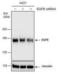
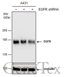
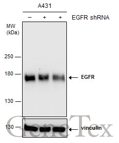
- Non-transfected (¡V) and transfected (+) A431 whole cell extracts (15 ?g) were separated by 5% SDS-PAGE, and the membrane was blotted with EGFR antibody [N1], N-term (GTX100725) diluted at 1:3000. The HRP-conjugated anti-rabbit IgG antibody (GTX213110-01) was used to detect the primary antibody.
Supportive validation
- Submitted by
- GeneTex (provider)
- Main image

- Experimental details
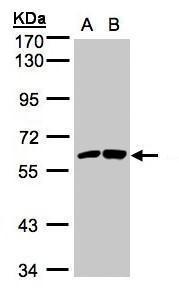
- Sample(30 ug whole cell lysate)A:H1299B:HeLa S3(GTX14654)7.5% SDS PAGEGTX100725 diluted at 1:1000
- Validation comment
- WB
- Submitted by
- GeneTex (provider)
- Main image

- Experimental details
- EGFR antibody [N1], N-term detects EGFR protein by western blot analysis.A. 30 ?g A431 whole cell extract7.5 % SDS-PAGEEGFR antibody [N1], N-term (GTX100725) dilution: 1:1000
- Validation comment
- WB

- Submitted by
- GeneTex (provider)
- Main image
- Experimental details
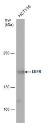
- EGFR antibody detects EGFR protein by western blot analysis. Whole cell extracts (30 ?g) was separated by 5% SDS-PAGE, and the membrane was blotted with EGFR antibody (GTX100725) diluted by 1:1000. The HRP-conjugated anti-rabbit IgG antibody (GTX213110-01) was used to detect the primary antibody.

- Submitted by
- GeneTex (provider)
- Main image
- Experimental details
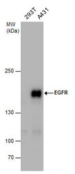
- EGFR antibody detects EGFR protein by western blot analysis. Whole cell extracts (30 ?g) was separated by 5% SDS-PAGE, and the membrane was blotted with EGFR antibody (GTX100725) at a dilution of 1:1000. The HRP-conjugated anti-rabbit IgG antibody (GTX213110-01) was used to detect the primary antibody.

- Submitted by
- GeneTex (provider)
- Main image

- Experimental details
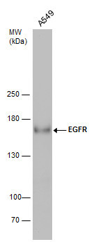
- Whole cell extract (30 ?g) was separated by 5% SDS-PAGE, and the membrane was blotted with EGFR antibody [N1], N-term (GTX100725) diluted at 1:500. The HRP-conjugated anti-rabbit IgG antibody (GTX213110-01) was used to detect the primary antibody.
- Submitted by
- GeneTex (provider)
- Main image
- Experimental details
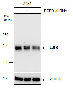
- Non-transfected (¡V) and transfected (+) A431 whole cell extracts (15 ?g) were separated by 5% SDS-PAGE, and the membrane was blotted with EGFR antibody [N1], N-term (GTX100725) diluted at 1:3000. The HRP-conjugated anti-rabbit IgG antibody (GTX213110-01) was used to detect the primary antibody.
Supportive validation
- Submitted by
- GeneTex (provider)
- Main image

- Experimental details
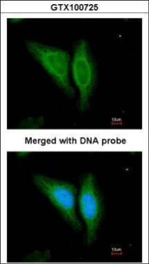
- Immunofluorescence analysis of paraformaldehyde-fixed HeLa, using EGFR(GTX100725) antibody at 1:200 dilution.
Supportive validation
- Submitted by
- GeneTex (provider)
- Main image

- Experimental details
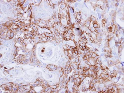
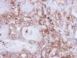
- Immunohistochemical analysis of paraffin-embedded human breast cancer, using EGFR(GTX100725) antibody at 1:100 dilution.

- Submitted by
- GeneTex (provider)
- Main image
- Experimental details
- Immunohistochemical analysis of paraffin-embedded CA922 xenograft, using EGFR(GTX100725) antibody at 1:500 dilution.