Explore
 Validate
 Learn
Learn Western blot
 Immunoprecipitation
Antibody data
- Antibody Data
- Antigen structure
- References [8]
- Comments [0]
- Validations
- Western blot [1]
- Immunocytochemistry [2]
- Other assay [2]
Submit
Validation data
Reference
Comment
Report error
- Product number
- AHR5072 - Provider product page

- Provider
- Invitrogen Antibodies
- Product name
- EGFR Monoclonal Antibody (199.12)
- Antibody type
- Monoclonal
- Antigen
- Purifed from natural sources
- Description
- AHR5072 reacts with an extracellular domain of EGFR. The molecular weight of the antigen is 170kDa (wild type) and 145kDa (vIII variant). This antibody does not block the binding of EGF to EGFR.
- Reactivity
- Human
- Host
- Mouse
- Isotype
- IgG
- Antibody clone number
- 199.12
- Vial size
- 500 µL
- Concentration
- 0.2 mg/mL
- Storage
- 4° C
Submitted references Tyrosine Phosphorylation of the Myosin Regulatory Light Chain Controls Non-muscle Myosin II Assembly and Function in Migrating Cells.
High CD90 (THY-1) expression positively correlates with cell transformation and worse prognosis in basal-like breast cancer tumors.
Direct visualization of single-molecule membrane protein interactions in living cells.
Ikarugamycin: A Natural Product Inhibitor of Clathrin-Mediated Endocytosis.
EGFR Overexpressed in Colonic Neoplasia Can be Detected on Wide-Field Endoscopic Imaging.
Differential effects of EGFR ligands on endocytic sorting of the receptor.
Sulindac metabolites inhibit epidermal growth factor receptor activation and expression.
Adhesion-mediated squamous cell carcinoma survival through ligand-independent activation of epidermal growth factor receptor.
Aguilar-Cuenca R, Llorente-González C, Chapman JR, Talayero VC, Garrido-Casado M, Delgado-Arévalo C, Millán-Salanova M, Shabanowitz J, Hunt DF, Sellers JR, Heissler SM, Vicente-Manzanares M
Current biology : CB 2020 Jul 6;30(13):2446-2458.e6
Current biology : CB 2020 Jul 6;30(13):2446-2458.e6
High CD90 (THY-1) expression positively correlates with cell transformation and worse prognosis in basal-like breast cancer tumors.
Lobba ARM, Carreira ACO, Cerqueira OLD, Fujita A, DeOcesano-Pereira C, Osorio CAB, Soares FA, Rameshwar P, Sogayar MC
PloS one 2018;13(6):e0199254
PloS one 2018;13(6):e0199254
Direct visualization of single-molecule membrane protein interactions in living cells.
Kim DH, Park S, Kim DK, Jeong MG, Noh J, Kwon Y, Zhou K, Lee NK, Ryu SH
PLoS biology 2018 Dec;16(12):e2006660
PLoS biology 2018 Dec;16(12):e2006660
Ikarugamycin: A Natural Product Inhibitor of Clathrin-Mediated Endocytosis.
Elkin SR, Oswald NW, Reed DK, Mettlen M, MacMillan JB, Schmid SL
Traffic (Copenhagen, Denmark) 2016 Oct;17(10):1139-49
Traffic (Copenhagen, Denmark) 2016 Oct;17(10):1139-49
EGFR Overexpressed in Colonic Neoplasia Can be Detected on Wide-Field Endoscopic Imaging.
Zhou J, Joshi BP, Duan X, Pant A, Qiu Z, Kuick R, Owens SR, Wang TD
Clinical and translational gastroenterology 2015 Jul 16;6(7):e101
Clinical and translational gastroenterology 2015 Jul 16;6(7):e101
Differential effects of EGFR ligands on endocytic sorting of the receptor.
Roepstorff K, Grandal MV, Henriksen L, Knudsen SL, Lerdrup M, Grøvdal L, Willumsen BM, van Deurs B
Traffic (Copenhagen, Denmark) 2009 Aug;10(8):1115-27
Traffic (Copenhagen, Denmark) 2009 Aug;10(8):1115-27
Sulindac metabolites inhibit epidermal growth factor receptor activation and expression.
Pangburn HA, Kraus H, Ahnen DJ, Rice PL
Journal of carcinogenesis 2005 Sep 2;4:16
Journal of carcinogenesis 2005 Sep 2;4:16
Adhesion-mediated squamous cell carcinoma survival through ligand-independent activation of epidermal growth factor receptor.
Shen X, Kramer RH
The American journal of pathology 2004 Oct;165(4):1315-29
The American journal of pathology 2004 Oct;165(4):1315-29
No comments: Submit comment
Supportive validation
- Submitted by
- Invitrogen Antibodies (provider)
- Main image
- Experimental details
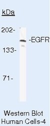
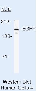
- Western blot analysis of EGFR in A431 cells using a EGFR monoclonal antibody (Product # AHR5072).
Supportive validation
- Submitted by
- Invitrogen Antibodies (provider)
- Main image

- Experimental details
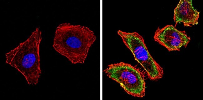
- Immunofluorescent analysis of EGFR (green) showing staining in the cytoplasm and membrane of HeLa cells. Formalin-fixed cells were permeabilized with 0.1% Triton X-100 in TBS for 5-10 minutes and blocked with 3% BSA-PBS for 30 minutes at room temperature. Cells were probed with a Epidermal Growth Factor Receptor monoclonal antibody (Product # AHR5072) in 3% BSA-PBS at a dilution of 1:50 and incubated overnight at 4 ºC in a humidified chamber. Cells were washed with PBST and incubated with a DyLight-conjugated secondary antibody in PBS at room temperature in the dark. F-actin (red) was stained with a fluorescent red phalloidin and nuclei (blue) were stained with Hoechst or DAPI. Images were taken at a magnification of 60x.
- Submitted by
- Invitrogen Antibodies (provider)
- Main image

- Experimental details
- Immunofluorescent analysis of EGFR (green) showing staining in the cytoplasm and membrane of A431 cells. Formalin-fixed cells were permeabilized with 0.1% Triton X-100 in TBS for 5-10 minutes and blocked with 3% BSA-PBS for 30 minutes at room temperature. Cells were probed with a Epidermal Growth Factor Receptor monoclonal antibody (Product # AHR5072) in 3% BSA-PBS at a dilution of 1:50 and incubated overnight at 4 ºC in a humidified chamber. Cells were washed with PBST and incubated with a DyLight-conjugated secondary antibody in PBS at room temperature in the dark. F-actin (red) was stained with a fluorescent red phalloidin and nuclei (blue) were stained with Hoechst or DAPI. Images were taken at a magnification of 60x.

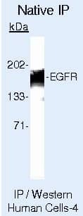
Supportive validation
- Submitted by
- Invitrogen Antibodies (provider)
- Main image

- Experimental details
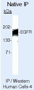
- Immunoprecipitation of EGFR in native human A431 cells using a EGFR monoclonal antibody (Product # AHR5072).
- Submitted by
- Invitrogen Antibodies (provider)
- Main image
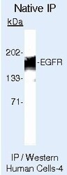
- Experimental details
- Immunoprecipitation of EGFR in native human A431 cells using a EGFR monoclonal antibody (Product # AHR5072).