 Explore
 Validate
 Learn
Learn Western blot
Antibody data
- Antibody Data
- Antigen structure
- References [3]
- Comments [0]
- Validations
- Western blot [1]
- Immunohistochemistry [1]
Submit
Validation data
Reference
Comment
Report error
- Product number
- PAB10009 - Provider product page

- Provider
- Abnova Corporation
- Proper citation
- Abnova Corporation Cat#PAB10009, RRID:AB_1707749
- Product name
- EGFR (phospho Y1197) polyclonal antibody
- Antibody type
- Polyclonal
- Description
- Rabbit polyclonal antibody raised against synthetic phosphopeptide of EGFR.
- Storage
- Store at 4°C. For long term storage store at -20°C.Aliquot to avoid repeated freezing and thawing.
Submitted references Role of epidermal growth factor receptor and STAT-3 activation in autonomous proliferation of SUM-102PT human breast cancer cells.
Expression of ras oncogene p21 protein in normal and neoplastic ovarian tissues: correlation with histopathologic features and receptors for estrogen, progesterone, and epidermal growth factor.
Occurrence of epidermal growth factor receptors in benign and malignant ovarian tumors and normal ovarian tissues: an immunohistochemical study.
Sartor CI, Dziubinski ML, Yu CL, Jove R, Ethier SP
Cancer research 1997 Mar 1;57(5):978-87
Cancer research 1997 Mar 1;57(5):978-87
Expression of ras oncogene p21 protein in normal and neoplastic ovarian tissues: correlation with histopathologic features and receptors for estrogen, progesterone, and epidermal growth factor.
Scambia G, Catozzi L, Panici PB, Ferrandina G, Coronetta F, Barozzi R, Baiocchi G, Uccelli L, Piffanelli A, Mancuso S
American journal of obstetrics and gynecology 1993 Jan;168(1 Pt 1):71-8
American journal of obstetrics and gynecology 1993 Jan;168(1 Pt 1):71-8
Occurrence of epidermal growth factor receptors in benign and malignant ovarian tumors and normal ovarian tissues: an immunohistochemical study.
Henzen-Logmans SC, van der Burg ME, Foekens JA, Berns PM, Brussée R, Fieret JH, Klijn JG, Chadha S, Rodenburg CJ
Journal of cancer research and clinical oncology 1992;118(4):303-7
Journal of cancer research and clinical oncology 1992;118(4):303-7
No comments: Submit comment
Supportive validation
- Submitted by
- Abnova Corporation (provider)
- Main image
- Experimental details
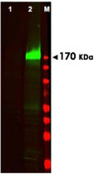
- Western blot using EGFR (phospho Y1197) polyclonal antibody (Cat # PAB10009) shows detection of a band at ~170 KDa corresponding to human EGFR (arrowhead).Staining is not seen in unstimulated A-431 cells (Lane 1), but is seen when A-431 cells are stimulated with EGF (50 ng/mL for 15 min) (lane 2). Approximately 30 ug of lysate was separated on a 4-20% Tris-Glycine gel by SDS-PAGE and transferred onto nitrocellulose.After blocking the membrane was probed with the primary antibody diluted to 1 : 250.Reaction occurred overnight at 4°C followed by washes and reaction with a 1 : 10,000 dilution of IRDye™800 conjugated Gt-a-Rabbit IgG [H&L] MX for 45 min at room temperature (800 nm channel, green).Molecular weight estimation was made by comparison to prestained MW markers in lane M (700 nm channel, red).IRDye™800 fluorescence image was captured using the Odyssey® Infrared Imaging System developed by LI-COR.IRDye is a trademark of LI-COR, Inc.

Supportive validation
- Submitted by
- Abnova Corporation (provider)
- Main image

- Experimental details
- Immunohistochemical staining of EGFR (phospho Y1197) polyclonal antibody (Cat # PAB10009) was used at 5 ug/mL to detect signal in a variety of tissues including multi-human, multi-brain and multi-cancer slides.This image shows faintly to moderately positive staining of placental trophoblasts at 40X. Tissue was formalin-fixed and paraffin embedded. The image shows localization of the antibody as the precipitated red signal, with a hematoxylin purple nuclear counterstain.Personal Communi-cation, Tina Roush, Life Span Biosciences, Seattle, WA.
- Validation comment
- Immunohistochemistry (Formalin/PFA-fixed paraffin-embedded sections)
