Explore
 Validate
 Learn
Learn Western blot
Antibody data
- Antibody Data
- Antigen structure
- References [3]
- Comments [0]
- Validations
- Western blot [1]
Submit
Validation data
Reference
Comment
Report error
- Product number
- PAB10010 - Provider product page

- Provider
- Abnova Corporation
- Proper citation
- Abnova Corporation Cat#PAB10010, RRID:AB_1674780
- Product name
- EGFR polyclonal antibody
- Antibody type
- Polyclonal
- Description
- Rabbit polyclonal antibody raised against synthetic peptide of EGFR.
- Storage
- Store at 4°C. For long term storage store at -20°C.Aliquot to avoid repeated freezing and thawing.
Submitted references The EGF receptor provides an essential survival signal for SOS-dependent skin tumor development.
Role of epidermal growth factor receptor and STAT-3 activation in autonomous proliferation of SUM-102PT human breast cancer cells.
Expression of ras oncogene p21 protein in normal and neoplastic ovarian tissues: correlation with histopathologic features and receptors for estrogen, progesterone, and epidermal growth factor.
Sibilia M, Fleischmann A, Behrens A, Stingl L, Carroll J, Watt FM, Schlessinger J, Wagner EF
Cell 2000 Jul 21;102(2):211-20
Cell 2000 Jul 21;102(2):211-20
Role of epidermal growth factor receptor and STAT-3 activation in autonomous proliferation of SUM-102PT human breast cancer cells.
Sartor CI, Dziubinski ML, Yu CL, Jove R, Ethier SP
Cancer research 1997 Mar 1;57(5):978-87
Cancer research 1997 Mar 1;57(5):978-87
Expression of ras oncogene p21 protein in normal and neoplastic ovarian tissues: correlation with histopathologic features and receptors for estrogen, progesterone, and epidermal growth factor.
Scambia G, Catozzi L, Panici PB, Ferrandina G, Coronetta F, Barozzi R, Baiocchi G, Uccelli L, Piffanelli A, Mancuso S
American journal of obstetrics and gynecology 1993 Jan;168(1 Pt 1):71-8
American journal of obstetrics and gynecology 1993 Jan;168(1 Pt 1):71-8
No comments: Submit comment
Supportive validation
- Submitted by
- Abnova Corporation (provider)
- Main image

- Experimental details
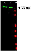
- Western blot using EGFR polyclonal antibody (Cat # PAB10010) shows detection of aband at ~170 KDa (Lane 1) corresponding to human EGFR present in unstimulated (Lane 1) and EGF (50 ng/mL for 15 min) stimulated (Lane 2) A-431whole cell lysates (arrowhead).Approximately 30 ug of lysate was separated on a 4-20% Tris-Glycine gel by SDS-PAGE and transferred onto nitrocell-ulose.After blocking the membrane was probedwith the primary antibody diluted to 1:1,000.Reaction occurred overnight at 4°C followed by washes and reaction with a 1 : 10,000 dilution of IRDye™800 conjugated Gt-a-Rabbit IgG [H&L] MX for 45 min at room temperature (800nm channel, green).Molecular weight estimationwas made by comparison to prestained MW markers in lane M (700 nm channel, red).IRDye™800 fluorescence image was captured usingthe Odyssey® Infrared Imaging System developedby LI-COR.IRDye is a trademark of LI-COR, Inc.